Learn about the comparison of reproductive system of various Arthropods.
Comparison: Arthropod # Macrobrachium:
1. The sexes are separate.
Male reproductive organs:
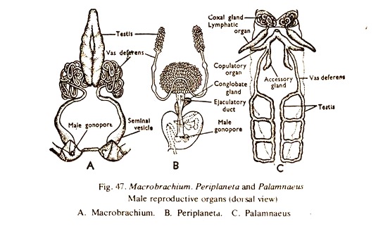
2. The male reproductive organs consist of a pair of testes, a pair of vasa deferentia, a pair of seminal vesicles and a pair of gonopores (Fig. 47A).
ADVERTISEMENTS:
3. The testes are soft, white, elongated bodies, fused at both the ends and are situated in the cephalothorax, below the heart and above the hepatopancreas.
4. From each testis arises a narrow tube, the vas deferens, which is much colied at first and then descends down towards the base of the fifth walking leg of the side.
5. The terminal end of each vas deferens forms a club-shaped swelling, known as seminal vesicle, which opens to the exterior by the male gonopore on the inner side of the coxa of the 5th walking leg.
Female reproductive organs:
ADVERTISEMENTS:
6. The female reproductive organs consist of a pair of ovaries, a pair of oviducts and a pair of female gonopores (Fig. 48A).
7. Ovaries are small and whitish in off-seasons but large and dark brown in the breeding season. They are fused at both the ends, larger in size than the testes and occupy same position as the testes in the male.
8. From the middle of the outer side of each ovary arises an oviduct, which narrows downwards to open in the gonopore on the inner side of the coxa of the 3rd walking leg of the side.
9. Fertilization is external and the development is direct.
Comparison: Arthropod # Periplaneta:
ADVERTISEMENTS:
1. The sexes are separate.
Male reproductive organs:
2. The male reproductive organs consist of a pair of testes, a pair of vasa deferentia, a pair of seminal vesicles, an ejaculatory duct, a conglobate gland and a gonopore (Fig. 47B).
3. The testes are small bodies made up of a number of vesicles and situated in the 4th and 5th abdominal segment below the terga.
4. Each testis is connected to a narrow duct, the vas deferens, which runs backwards, inwards and downwards.
5. Each vas deferens before uniting with its fellow of the opposite side dilates a little forming a seminal vesicle, from which arise a large number of blind pouches and the whole arrangement is known as mushroom gland.
After forming the structure, the two vasa deferentia unite and form a muscular tube, the ejaculatory duct, which opens in the gonopore, below the anus, guarded by go- napophyses. The conglobate gland is associated with the ejaculatory duct.
Female reproductive organs:
6. The female reproductive organs consist of a pair of ovaries, a pair of oviducts, a pair of colleterial glan.ds, a pair of spermathecae and a pair of gonopores (Fig. 48B).
ADVERTISEMENTS:
7. Ovaries are situated in the abdomen. Each ovary consists of 8 ovarioles connected anteriorly with the body wall by a ligament.
8. From the posterior end of the ovariole runs a narrow tubule and by the union of tubules coming from all the ovarioles of an ovary, a wider tube, the oviduct is formed. The two oviducts unite to form a median chamber, sometimes called the vagina, which opens in the gonopore guarded by the gonapophyses on the sternum of the 8th abdominal segment. A pair of asymmetrical spermathecae of which only one is functional, open together in the middle of the sternum of the 9th segment. A pair of colleterial glands opens behind the spermathecae.
9. Fertilization is internal. 16 eggs are enclosed in a capsule secreted by the colleterial glands. Development direct, with a nymph stage.
Comparison: Arthropod # Palamnaeus:
1. The sexes are separate.
Male reproductive organs:
2. The male reproductive organs consist of a pair of testes, a pair of vasa deferentia, a pair of genital chambers, a pair of paraxial organs, a pair of flagella, a pair of seminal vesicles, a common genital atrium and a gonopore (Fig. 47C).
3. Each testis consists of a pair of longitudinal tubules placed in the preabdomen, and connected by four cross branches.
4. The two longitudinal tubules of each testis unite anteriorly to form a vas deferens, which runs forward and downwards.
5. The terminal portion of each vas deferens opens into the inner side of the genital chamber along with an enlarged seminal vesicle. The genital chambers are muscular tubes and bear the paraxial organ having a chitinoid flagellum. It also receives the accessory gland. The genital chambers unite to form a median genital atrium, which opens to the exterior by the gonopore situated just behind the operculum.
Female reproductive organs:
6. The female reproductive organs consist of a single ovary, two oviducts, a median genital chamber and a female gonopore (Fig.48C).
7. Ovary is situated in the preabdomen. It consists of three longitudinal tubules connected by cross branches. The median tubule is shorter than the other two.
The ovarian tubules and the cross branches bear numerous diverticula.
8. From the two lateral tubules run two narrow oviducts. The two oviducts open in the sac-like median genital chamber, the later opening in the gonopore on the second abdominal segment below the bilobed operculum.
9. Fertilization is internal. Development is direct. The embryos develop in the diverticula of the tubules and the young ones are born (viviparous).
