The following points highlight the four main types of classification of placenta. The types are: 1. Classification Based on the Degree of Intimacy 2. Classification Based on the Types of Implantation 3. Classification Based on the Distribution of Villi 4. Classification Based on the Degree of Involvement of Foetal and Maternal Tissues.
Classification of Placenta:
- Classification Based on the Degree of Intimacy
- Classification Based on the Types of Implantation
- Classification Based on the Distribution of Villi
- Classification Based on the Degree of Involvement of Foetal and Maternal Tissues
Type # 1. Classification Based on the Degree of Intimacy:
Based on the degree of intimacy of foetal and maternal tissues, the following types of placenta are seen:
ADVERTISEMENTS:
i. Non-Deciduous Placenta or Semi Placenta:
In non-deciduous placenta the implantation is superficial. This occurs in most mammals where the blastocysts lie in the uterine cavity. At the point of contact with the wall of the uterus, the blastocyst surface gives out finger like projection called chorionic villi that penetrate into the depressions of the uterine wall and are loosely united.
At the time of birth, when parturition takes place, the chorionic villi are simply withdrawn from the cavities of the uterine wall without causing any damage or bleeding. This type of placenta formation is seen in pig, cattle, horse etc., where the less hazardous birth process allows the females to resume full running speed soon after birth.
As the chorionic villi do not fuse with the endometrium, such a placenta is also called semi placenta.
ADVERTISEMENTS:
ii. Deciduous Placenta or Placenta Vera:
In cat, dog, primates, rodents etc., the degree of intimacy between the chorionic villi and the endometrium is greatly increased. The uterine wall gets eroded. The chorionic villi fuse with the eroded uterine mucosa. Such a placenta is termed as placenta vera (true placenta).
At the time when parturition takes place the uterine wall does not remain intact. It tears away and extensive haemorrhage takes place at birth. Such a type of placenta is termed as deciduous placenta.
This phenomena of shedding (tearing off) and replacement of maternal tissue is termed as decidua (meaning, to shed). Here the placenta is physiologically more efficient, where the mothers are protected enough to recover fully after child birth.
ADVERTISEMENTS:
iii. Contra-Deciduate Placenta:
A somewhat modified type of deciduate placenta is seen in Parameles and Talpa (mole), where there is loss of both maternal tissue as well as foetal portion of placenta. Such a placenta is called contra-deciduate placenta.
Type # 2. Classification Based on the Types of Implantation:
Implantation is the process by which the embryo becomes attached to a nutritional substance. The term in case of placental mammals is referred to the process by which the embryo remains associated intimately with the uterine wall.
Generally three types of implantation are seen which are as follows:
i. Central or Superficial Implantation:
The chorionic sac of the embryo grows and makes superficial attachment with the uterine mucosa. This type of implantation is called central or superficial implantation and the embryo remains within the lumen of the uterus (Fig. 5.53A).
It is seen in all cases of implantation in lower vertebrates. It is also present in Preambles and Dasyurus among the marsupials, while among eutherians it is seen in pig, cow, rabbit, sheep, dog, cat etc.
ii. Eccentric Implantation:
ADVERTISEMENTS:
In mouse, rat, beaver, squirrel etc., the blastocyst in its early stage comes to lie between the uterine epithelial folds or pocket (Fig. 5.53B), and this type of implantation is called eccentric implantation. The epithelial folds at a later stage, encloses the blastocyst almost completely.
iii. Interstitial Implantation:
In interstitial implantation the embryo burrows into the uterine mucosa below the epithelium and becomes surrounded completely by the endometrial tissue of the uterus (Fig. 5.53C). This type of implantation is seen in hedgehog, guinea-pig, some bats, chimpanzee, man etc.
Type # 3. Classification Based on the Distribution of Villi:
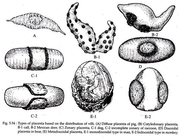
The pattern of distribution of villi varies among species of different mammals. Based on this the following types of placenta are recognized (Fig. 5.54).
i. Diffused Placenta:
In diffused placenta the villi are numerous and are scattered uniformly over the whole of chorion. It is seen in ungulates (pig, horse, mare etc.) and in cetacea.
ii. Cotyledonary Placenta:
In this type of placenta the villi become aggregated in special regions or patches to form small tufts. The rest part of the chorion surface remains smooth. It is seen in ruminant (cud-chewing) ungulates such as cattle, sheep, deer etc. In camel and giraffe an intermediate type of placenta is seen where the villi are scattered and are also arranged in cotyledons.
iii. Zonary Placenta:
In a zonary placenta the villi are confined to an annular or girdle-like zone on the chorion (chorion is more or less elliptical in shape). Such a placenta occurs in carnivores and may be of either incomplete zonary (e.g. raccoon) or complete zonary (e.g. dog, cat, seal etc.) type.
iv. Discoidal Placenta:
Here the villi become restricted to a circular disc or plate area on the dorsal surface of blastocyst. Such a placenta is seen in insectivores, bats, rodents (rat, mouse), rabbit and bear.
v. Meta-Discoidal Placenta:
In primates a special type of discoidal placenta is seen where the villi are at first scattered all over the chorion but later becomes restricted to one or two discs. The mono-discoidal type with a single disc is seen in man, while the bi-discoidal type with two disc shaped villous areas is seen in monkeys.
Type # 4. Classification Based on the Degree of Involvement of Foetal and Maternal Tissues:
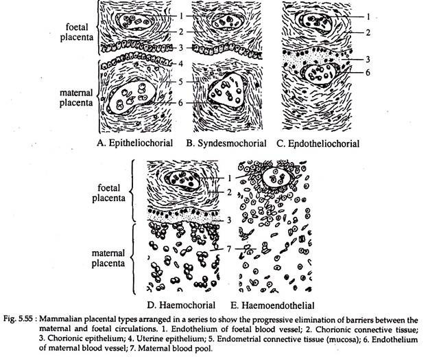
Histologically there are no less than six membranes or tissues (called barriers) that lie between the foetal and maternal blood streams. These membranes in order of their sequence, from mother to foetus are; endothelium of maternal blood vessel, endometrial connective tissue, uterine epithelium, chorionic epithelium, chorionic connective tissue and endothelium of foetal blood vessel.
The presence or absence of these tissues results in the formation of the following types of placenta (Fig. 5.55).
i. Epitheliochorial Placenta:
This type of placenta involves the contact of the chorionic epithelium with that of uterine epithelium and thus the term epitheliochorial. The epitheliochorial placenta involves six tissue barriers between the foetal and maternal circulation. It is the primitive type from which others have been derived and is seen in pig, sow, mare, horse, cattle etc.
ii. Syndesmochorial Placenta:
In the ruminant ungulates (cattle, sheep, deer, giraffe etc.), varying amounts of the uterine epithelium may be absent. As a result the chorion is brought into direct contact with the connective tissue of the uterus. Only five barriers therefore, lie between the two blood streams. This type of placenta is termed as syndesmochorial.
iii. Endotheliochorial Placenta:
In endotheliochorial placenta the uterine mucosa is reduced and the chorionic epithelium comes in contact with the endothelial wall of maternal blood vessels. This type of placenta is characteristic of carnivores like dog, cat, bear etc. Here, only four barriers remain between the two blood streams.
iv. Haemochorial Placenta:
A reduction of the barriers into three is observed in haemochorial placenta, which is found in primates, insectivores (mole, shrew) and chiropterans (bat). Here, the endothelial wall of the maternal blood vessel disappears and the chorionic epithelium is bathed directly in the maternal blood.
v. Haemoendothelial Placenta:
In this type of placenta the chorionic villi looses their epithelium and mesenchymal layers to such a degree that the endothelial wall of the foetal blood vessels remain in contact with the maternal blood. The haemoendothelial placenta is observed in mouse, rat, rabbit, guinea-pig etc. and only a single barrier separates the two blood streams.


