Learn about the comparison of urinogenital systems in various vertebrates.
Comparison: Vertebrates # Bufo:
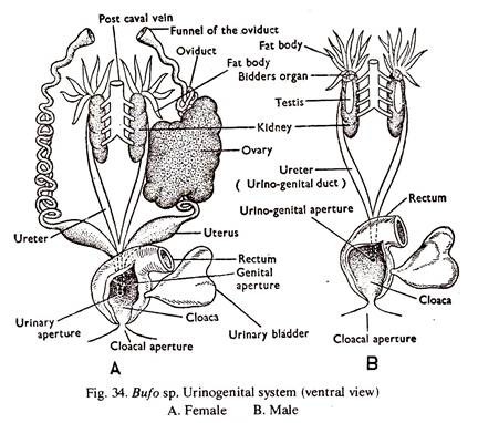
A. Urinary System – It consists of a pair of kidneys, a pair of ureters, a bilobed urinary bladder rand a urinary opening.
1. The kidneys are flat, elongated, dark-red lobulated structures situated in the coelom, dorsally on either side of the vertebral column. The adult kidney is mesonephric.
2. From the outer side of each kidney arises a white tube, the ureter. The ureter gradually enlarges, runs backward and the two unite posteriorly to open into the cloacal chamber on its dorsal surface. A thin-walled bilobed structure, the urinary bladder, in which urine is stored, opens into the cloacal chamber. The cloaca opens to the exterior through the cloacal aperture.
ADVERTISEMENTS:
B. Genitals system (Fig. 34) – The individual is either male or female.
Male genital system – It consists of a pair of tests, vasa efferentia, two urinogential ducts and their openings into the cloaca.
1. The testes are two, elongated, cream-white bodies attached to the ventral surface of the kidnes by means of a membraneos fold, known as mesorchium.
2. Several fine ducts called vasa efferentia pass from the testis to the kidney and finally open into the ureter.
ADVERTISEMENTS:
3. The ureter and the urinary opening serve as genital duct and its opening.
4. The spermatozoa produced in the testes pass to the ureters through the vasa efferentia and from there to the exterior through the common urinogenital opening and the cloacal aperture.
Female genital system – It consists of a pair of ovaries, a pair of oviducts with their funnels, a pair of uteri and a genital opening.
1. The ovaries are attached to the ventral surface of the kidneys by means of a membraneous fold, known as mesovarium. Ovaries are small and light brown in off seasons but large and with small black and white rounded structures during breeding season.
ADVERTISEMENTS:
2. Very near the heart, on each side, is a funnel like structure, the infundibulum. It runs posteriorly as a narrow, much-coiled tube, the oviduct (Mullerian duct). Unlike the male, the genital duct is separate from the ureter. The oviduct dilates posteriorly and forms the uterus. The two uteri unite posteriorly and opens dorsally into the cloaca.
3. The ova escape into the coelom by the rupture of the ovarian wall. The ova enter the oviduct through the infundibulum and receive a coating of albumen while passing downward. The eggs are discharged on water at the time of mating.
Comparison: Vertebrates # Calotes (Fig. 35. A,B):
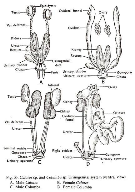
A. Urinary system – It consists of a pair of kidneys, a pair ureter, a urinary bladder and two urinary apertures.
1. The kidneys are flat, medium-sized, dark-red bodies with two lobes, situated in the posterior region of the coelom on either side of the vertebral column. The posterior ends of the kidneys are tapering and in close contact with each other. The adult kidney is metanephric.
2. From the outer side of each kidney arises a white tube, the ureter. The two ureters run backward and open dorsally into the cloaca by separate urinary apertures. A thin-walled sac-like structure, the urinary bladder, in which urine is stored, opens into the cloaca on its ventral surface. The colaca opens to the exterior by a cloacal aperture. In male, the ureter joins the vas deferens posteriorly to form a urinogenital duct which opens into the cloaca.
B. Genital system (Fig.35B) – The individual is either male or female.
Male genital system – Its consists of a pair of testes, two epididymis, two vasadeferentia, two copulatory organs and two urinogenital openings.
1. The testes are two, oval, white bodies attached to the dorsal body wall by the mesorchium. The right testis is slightly anteriorly placed.
2. From the inner side of each testis arises a narrow convoluted duct, the epididymis, which runs backward.
ADVERTISEMENTS:
3. The epididymis passes behind into a narrow convoluted duct, the vas deferens, which opens into the terminal part of the corresponding ureter. The rest of the ureter serves as a common urinogenital duct. A pair of copulatorysacs with bifid apex open into the posterior part of the cloaca.
4. The spermatozoa produced in the testes pass through the epididymis, vasa deferentia, urinogenital ducts and reach the cloaca. During mating, the spermatozoa come out through the hemipenis which is in the form of a pair of eversible sacs.
Female genital system – It consists of a pair of ovaries, a pair of oviducts with their funnels, and a pair of female genital apertures
1. The ovaries are attached to the dorsal body wall in the posterior region of the coelom by means of a membraneous fold, known as mesovarium. Ovaries are irregularly oval bodies with surfaces raised up into rounded elevations.
2. Much anterior to the ovaries is a pair of flat thin-walled tubes, the oviducts. The anterior end of each tube opens by a funnel-shaped structure, the infundibulum. Posteriorly, each oviduct is differentiated into a middle portion known as uterus and a terminal portion, the vagina, which opens into the posterior part of the cloacal chamber by a separate aperture. The oviducts are attached to the body wall by ligaments.
3. The ova escape into the coelom by the rupture of the ovarian wall. The ova enter the oviduct through the infundibulum and receive a coating of albumen while passing downward. It further receives a coating of calcium carbonate after fertilization and the eggs are laid on land.
Comparison: Vertebrates # Columba (Fig. 35 C,D):
A. Urinary system – It consists of a pair of kidneys, a pair of ureters and a pair of urinary apertures.
1. The kidneys are flat, elongated, dark-red bodies with three lobes. They closely fit into the follows of the pelvis. The adult kidney is metanephric.
2. From the ventral surface of each kidney arises a narrow duct, the ureter. The two ureters run straight backward and open directly into the middle chamber or urodaeum of the cloaca by separate urinary apertures.
B. Genital System (Fig. 35. C,D) – The individual is either male or female.
Male genital system – It consists a pair of testes, a pair of vasa deferentia, two vesiculae seminalis and two male genital apertures.
1. The testes are ovoid, white bodies, attached to the ventral surface of the anterior ends of the kidneys by the peritoneum.
2. From the inner border of each testis arises a narrow convoluted duct, the vas deferens which runs backward, parallel to the ureter.
3. Posteriorly, the vas deferens enlarges slightly and forms the vesicular seminalis. It opens into the urodaeum on the extremity of a small papilla.
4. The spermatozoa produced in the testes pass through the vasa deferentia and are stored temporarily in the vesiculae seminalis. Finally the spermatozoa pass out through the cloacal aperture during mating.
Female genital system – It consists of an ovary, an oviduct situated on the left side, the right counterparts being atrophied, leaving a trace only, and a female genital opening.
1. The left ovary is attached to the dorsal body wall of the posterior region of the coelom by means of a membraneous fold, known as mesovarium. The ovary is irregularly oval with the surface raised up into rounded elevations due to the presence of follicles.
2. The left oviduct is along and convoluted tube, provided with a large funnel or infundibulum at the anterior end near the ovary. The wall of the tube is muscular in its middle region. The posterior portion is known as uterus, which is glandular and opens into the urodaeum of the cloaca by the genital aperture. The right oviduct is atrophied and a vestige persists.
3. The ova escape into the coelom by the rupture of the follicles and enter the oviduct through the infundibulum. While passing downward, each ovum receives albumen. After fertilization, it further receives a coating of double shell membrane and a calcareous shell, and the eggs are laid in nests.
Comparison: Vertebrates # Cavia (Fig. 36):
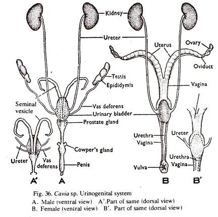
A. Urinary system – It consists of a pair of kidneys, a pair of ureters, a urinary bladder and urethra and a urinary aperture.
1. The kidneys are two, dark-red, bean-shaped bodies attached to the dorsal body wall by the sides of the vertebral column. The right one is slightly anteriorly placed than the left. Each kidney is composed of two parts, the outer cortex and the inner medulla. The adult kidney is metanephric.
2. From the inner concave region or hilum of the kidney arises a narrow duct, the ureter which runs backward and the two ureters open separately into the urinary bladder on either lateral side. The urinary bladder is a thin-walled median sac, the neck of which is drawn into a narrow tube, the urethra.
In the male, the urethra traverses the penis and opens to the exterior at its tip. In the female, it is small and opens to the exterior by a small urinary aperture behind the clitoris.
B. Genital system (Fig. 36). The individual is either male or female.
Male genital system – It consists of a pair of tests, two epididymis, two vasa deferentia, two seminal vesicles, a penis, certain glands and the genital opening.
1. The testes are white ovoid bodies situated in the coelom in the off seasons, but descends into the scrotum during breeding season.
2. From each testis arises a much convoluted tubule, the epididymis which remains attached to the testis.
3. The edididymis runs as vas deferens and the two vasa deferentia open into the neck of the urinary bladder by a common aperture on is dorsal surface. The penis is an elongated, muscular, rounded structure, the tip of which is known as glans penis. The junction of the urethra, spermiduct and ureter is surrounded by a large gland the prostate gland. A paired diverticulum, the vesciculae seminalis are situated close to the urinary bladder.
4. The spermatozoa produced in the testes pass through the epididymis and the vasa deferentia and reach the seminal vesicle. During mating the spermatozoa pass to the exterior through the common urinogenital opening at the tip of the penis.
Female genital system – It consists of a pair of ovaries, two infundibula, two oviducts, a vagina, a vestibule and vulva.
1. The ovaries are small and attached to the dorsal body wall of the posterior part of the abdomen, by the sides of the backbone by means of a membraneous fold, known as mesovarium.
2. Very near each ovary is a funnel-shaped structure, the infundibulum, which is connected posteriorly with the oviduct. The first part of the oviduct is very narrow, coiled and known as fallopian tube. The middle part is wide and called uterus. The two uteri unite posteriorly and is known as vagina. The posterior part of the vagina, the vestibule, opens to the exterior through the female genital aperture or vulva. Two prominent folds known as labia majora are situated by the side of the vulva. A small muscular structure, the clitoris, homologous to the penis of the male is situated in front of the urinary opening.
3. The ova escape into the coelom by the rupture of the ovarian wall and enter the oviduct through the infundibulum and come to rest in the fallopian tube. During mating, spermatozoa discharged into the vagina move forward and fertilize the ova. The zygotes come to the uteri where development proceeds and nourishment is drawn through the placenta. Young individuals are born directly.


