Learn about the comparison of hind limb bones in various vertebrates.
Comparison: Vertebrates # Bufo (Fig. 27 B):
1. The bones of the hind limb consist of a femur, a tibiofibula, tarsal and metatarsal bones and phalanges.
2. Femur. It has a long, narrow, rounded shaft. The proximal end bears a rounded structure known as the head which fits into the acetabulum of the pelvic girdle. The distal end bears a process for articulation with the tibiofibula.
3. Tibiofibula. It has a long shaft, narrow and round at the middle but broad and flat at the ends. It is formed by the fusion of two bones, the tibia and the fibula. The tibial portion bears a slight ridge, the cnemial crest, towards the proximal end.
ADVERTISEMENTS:
4. Tarsal bones. These are the bones of the ankle and arranged in two rows. The proximal tarsals are two in number, elongated and united at both the ends. The curved one is astragalus and the straight one, the calcanemu. The distal tarsals are two and small in sizes.
5. Metatarsal bones. Threse are the bones of the foot and five in umber corresponding to five fingers.
6. Phalanges. These are the bones of the fingers. The number of phalanges in each finger from first to fourth are 2,2, 3 and 3 respectively.
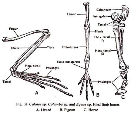
Comparison: Vertebrates # Calotes (Fig. 32 A):
1. The bones of the hind limb consist of a femur, a tibia and fibula, tarsal and metatarsal bones and phalanges.
ADVERTISEMENTS:
2. Femur. It has a long and stout shaft with epiphysis at both the ends. The proximal end bears a rounded head which fits into the acetabulum of the pelvic gridle. Near the head is a rought-surfaced trochanter. The distal end bears two ballshaped condyles for articulation with the tibia and a tuberosily for the fibula.
3. Tibia and Fibula. It consists of two elongated separate bone, the tibia and the fibula. The tibia is a stout, curved bone and bears a cnemial crest. The proximal end bears two articular surfaces for the femur and the distal end has a concavity. The fibula is slender bone with an articular surface at the proximal end. The distal end is convex.
4. Tarsal bones. These are the bones of the ankle and three in number. The large proximal one is known as tibiofibulare and the two distals are smaller.
5. Metatarsal bones. These are the bones of the foot, five in umber corresponding to the five fingers.
ADVERTISEMENTS:
6. Phalanges. These are the bones of the fingers. The number of phalanges in each finger from first to fifth are 2,3,4 and 5 respectively.
Comparison: Vertebrates # Columba (Fig. 32 B):
1. The bones of the hind limb consist of a femur, a tibiotarsus and fibulam a tarsometatarsus and the phalanges.
2. Femur. It has a long, cylindrical and stout shaft. The proximal end bears a rounded head at right angle to the shaft, which fits into the acetabulum of the pelvic girdle. Near the head and at right angle to the shaft is trochanter. The distal end is pulley-like and the condyles are separated by intercondylar fossa. A patella is present at the knee joint.
3. Tibiotarsus and fibula. The tibiotarsus is a long bone with a cnemial process near the proximal end. The proximal end bears a concave articular surface and the distal end is pulley-like. The fibula is a very small bone which ends to a point at the distal end. The tarsal bones (astragalas and calcaneum) fuse with the tibia to form the tibiotarsus.
4. Trasometatarsus. It is an elongated bone, the proximal end bears a concave surface and the distal end three distinct pulleys for the articulation of three toes. It is formed by the fusion of the distal tarsals and three metatarsals.
5. Metatarsal bones. Only the first metatarsal is a free bone. It is of irregular shape and attached near the distal end of the tarsome tatarsus.
6. Phalanges. These are the bones of the fingers. The number of the phalanges in each finger from first to fourth are 2,3, 4 and 5; in all cases the last phalanx ends in a claw.
Comparison: Vertebrates # Cavia (Fig. 29 B):
1. The bones of the hind limb consist of a femur, a tibia andfibula, tarsal and metatarsal bones and phalanges.
2. Femur. It has a long, stout, slightly curved shaft. The proximal end bears a prominent head to fit into the acetabulum of the pelvic girdle. Three elevated areas, greater trochanter immediately below the head and lesser and third trochanter below the greater trochanter are present. The distal end is pulley like. The condyles are separated by an intercondylar notch. A patella is present at the knee joint.
ADVERTISEMENTS:
3. Tibia and fibula. It consists of two separate bones attached at the ends only. The tibia is strong, stouter at the anterior end and narrower towards the posterior end. Two concave facets are present at the proximal end. A cnemial crest is present towards the proximal end. The distal end also bears concavity. The fibula is slender and narrows towards the distal end.
4. Tarsal bones. These are the bones of the ankle and six in number. The proximal two are large and known as astragalus and calcaneum. The distal tarsals are small in size.
5. Metatarsal bones. These are the bones of the foot, three in number corresponding to the three fingers.
6. Phalanges. These are the bones of the fingers. The number of phalanges in each finger is three, the distal one ending in a claw.
Comparison: Vertebrates # Equus (Fig. 32 C):
1. The bones of the hind limb consist of a femur, a tibia and fibula, tarsal and metatarsal bones and phalanges.
2. Femur. It is like that of the cavia.
3. Tibia and fibula. It is more or less like that of the cavia.
4. Tarsal bones. These are the bones of the ankle and six in number. The astragalus has a pulley-like surface above for articulation with the tibia. Its distal surface is flattened and articulates more with the navicular than with the cuboid. The calcaneum does not articulate with the fibula. The fused meso- ento- and ecto- cuneiform articulate distally with the metatarsals.
5. Metatarsal bones. These are the bones of the foot. The first and fifth are completely degenerated. The second and fourth are represented by narrow splint a large, elongated, round bone known as cannon bone.
6. Phalanges. These are the bones of the fingers. The 2nd and 4th fingers are represented by small bones, the sesamoids. The third finger is elongated and consists of three phalanges, the last one bearing a hoof.
