
The below mentioned article provides a short note on the Placentation in Rabbit:- 1. Meaning of Placentation in Rabbit 2. Process of Placentation 3. Organogenesis of Placenta in Rabbit.
Meaning of Placentation in Rabbit:
The egg of rabbit after fertilization reaches the uterus which consists of two long, tubular “horns”. A single cleaving egg or blastocyst generally lodges itself in one of the crypts at the midpoint of the horn containing it.
If two or more cleaving eggs are present, they become evenly spaced along the horn. To prevent the sticking of the blastocyst with any other maternal tissue, the zona pellucida remains attached to it, till it reaches the uterine cavity.
As soon as fertilization is completed the corona radiata cells and zona pellucida breaks down to form a frothy substance called histotroph. The histotroph surrounds the entire fertilized egg and provides nutrition for its further growth and development.
ADVERTISEMENTS:
The egg grows vigorously at the expense of the histotroph and soon completes the morphogenetic movements to produce the three primary germinal layers — endoderm, mesoderm and ectoderm.
Process of Placentation:
After the three primary germ layers are laid down in the embryo, remarkable physiological changes take place in the uterine wall of the mother. The region where the uterine wall comes in contact with the embryo, bleeds due to some enzymatic action.
The process of implantation in rabbit is initiated and controlled by a protein called blastokinin which is secreted by the uterine wall. The maternal site of implantation subsequently becomes thick, spongy and vascularized. The superficial contact is made intimate by the formation of minute fingerlike projections called villi (Fig. 5.64).
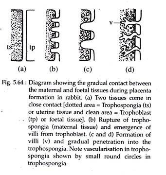
The villi develop as an outgrowth from the extra embryonic region of the foetus and penetrate into the depressions present in the wall of the uterus. These villi are initially formed by the trophoblast. Later the connective tissue and blood vessels enter into it. At this time they are designated as “chorionic villi” and the blood vessels that enter into it are the ramifications of the allantoic blood vessels.
ADVERTISEMENTS:
The portion of the uterine wall that has undergone vascularization is called trophospongia. The villi of the trophoblast penetrate deep into the trophospongia and ramify extensively into it, establishing intimate physiological relationships. During this action, the villi are constantly bathed by the maternal blood due to excessive vascular state of the trophospongia.
The placenta, thus, is represented jointly by the chorion and villi of the trophoblast and the endometrium of the trophospongia. After forming close physiological union, the trophoblast and trophospongia form a nutritional bridge, where the maternal blood brings along with it nutritional materials for the developing embryo throughout the period of gestation.
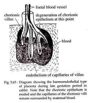
In rabbit, the epithelium of the chorion may disappear in some regions during the later stages of gestation, thereby permitting an exposure of foetal blood vessels to the maternal blood (Fig. 5.65).
Organogenesis of Placenta in Rabbit:
In rabbit the initial contact of the embryonic tissue with the uterine wall forms an epitheliochorial relationship. Subsequently in course of development, the relationship changes into a haemochorial condition in which the endothelial tissue of the uterine blood vessels is destroyed by the erosive action of the foetal tissue.
This results into the chorionic epithelium of the embryonic part of the placenta to come into direct contact with the maternal blood. During later stages of pregnancy, even the chorionic epithelium disappears, leaving the endothelial lining of the foetal blood vessels in direct contact with the maternal blood.
This type of placental relationship is called haemoendothelial type (Fig. 5.65), and is regarded to be the most intimate grade of placental relationship in the animal kingdom.
Further, depending upon the distribution of villi, the placenta of rabbit is categorized as the discoidal type. At the initial stage of placentation the chorion becomes uniformly covered with villi, but at a later stage the villi remain only on one side.
The villi close to the uterine wall become well developed and form a disc shaped structure, while the villi on other parts of the chorion become atrophied. At the time of parturition in rabbit the chorionic villi are simply drawn out from the crypts of the uterine wall.