
The below mentioned article provides an overview on Periplaneta (Cockroach):- 1. Introduction to Periplaneta 2. Habit and Habitat of Periplaneta 3. Structure 4. Integumentary System 5. Muscular System 6. Body Cavity 7. Locomotion 8. Digestive System 9. Respiratory System 10. Circulatory System 11. Excretory System 12. Nervous System 13. Endocrine System 14. Reproductive System.
Contents:
- Introduction to Periplaneta
- Habit and Habitat of Periplaneta
- Structure of Periplaneta
- Integumentary System of Periplaneta
- Muscular System of Periplaneta
- Body Cavity of Periplaneta
- Locomotion of Periplaneta
- Digestive System of Periplaneta
- Respiratory System of Periplaneta
- Circulatory System of Periplaneta
- Excretory System of Periplaneta
- Nervous System of Periplaneta
- Endocrine System of Periplaneta
- Reproductive System of Periplaneta
1. Introduction to Periplaneta:
Cockroaches are common household pests and also act as a scavenger. The word cockroach has originated from the name of a Spanish fruit ‘cucaracha’, having disagreable taste.
ADVERTISEMENTS:
Its four common species found in India are:
(i) Blatella germanica, the German cockroach,
(ii) Blatta orientalis, the Oriental cockroach,
(iii) Periplaneta americana, the American cockroach and
ADVERTISEMENTS:
(iv) Periplaneta australasiae, the Australian cockroach.
Different species of cockroaches differ in size and other characteristics, but all of them exhibit certain common features. These are:
(i) Well developed wings present in both sexes,
(ii) The wings in males extend beyond the abdomen and
ADVERTISEMENTS:
(iii) In females keel-like ovipositor valves are present. The species described in this text is Periplaneta americana.
The generic name Periplaneta was assigned to it by Burmeister in 1838. It has become well established throughout India.
Etymology:
Greek: Peri, about; planeta, wandering star or planet. It indicates the worldwide distribution of this insect.
2. Habit and Habitat of Periplaneta:
Cockroach is a common nocturnal omnivorous scavenger. It prefers dark warm corners of kitchen, godowns, underground drains and places where food and humid atmospheres are available. Their narrow and flattened body enables them to slip into narrow creeks and crevices. Cockroaches are now distributed throughout the world.
They probably originated in Southern Asia or Africa and became the dominant fossils of the carboniferous age. Being omnivorous and scavengers they devour any kind of food and even the dead bodies of some animals. Cannibalism is often seen among cockroaches. Their ability to walk rapidly and the production of a pungent secretion from their abdominal glands has been regarded as their defensive mechanism.
3. Structure of Periplaneta:
The body of cockroach is dorsoventrally flattened, elongated and reddish-brown in colour. The males are usually 35-40 mm in length while that of females are 29-37 mm.
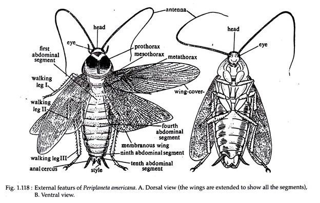
Body of Periplaneta is segmented (Fig. 1.118) and are organised to form three distinct specialised parts, each of which is called a tagma. The three tagmata are — head, thorax and abdomen. All of these are enclosed by exoskeleton. Each tagma comprises of several structures.
A. Head:
ADVERTISEMENTS:
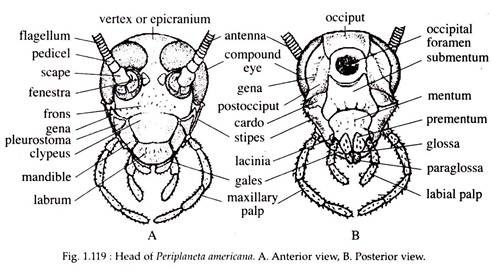
Head is the anterior most part of the body and is more or less triangular in shape. It is formed by the fusion of six segments which have lost their external demarcation. The exoskeleton covering the head is called the head capsule and it includes five sclerites (Fig. 1.119).
They are:
(i) A pair of epicranial plates or occipital sclerites, covering dorsal and posterior parts, and joined in front by an inverted y-shaped coronal or epicranial suture;
(ii) A single piece formed by the fusion of two exoskeletons frons and clypeus, the latter forming the lower part of the face and
(iii) The two sides of the head are covered by two vertical plates called the cheeks or genae, lying below the eyes. On each side the gena is separated from the frons by a frontogenal suture and the labrum is attached with the distal border of the clypeus.
The head region bears the following structures:
(a) Eye:
One on each side of the head, a pair of prominent sessile eyes are present. The two eyes occupy much of the anterolateral wall of the head. Each eye is bean-shaped and compound in nature. The external surface is marked with several polygonal facets, each of which denotes a single visual unit, the ommatidium. The eyes are larger in males than in females.
(b) Antenna:
The antennae are a pair of long, jointed, thread-like, movable structures present in the anteromedial indentation of the eyes. Each antenna articulates on a ring-like sclerite at two points — one, which is rigid is known as antennifer and the other is flexible, called surantennifer. The antennae can be moved freely in all directions.
(c) Ocellus:
The ocelli are paired circular areas one on each side of the eye, near the base of the antennae. Each area acts as a simple eye and is responsible for detecting light,
(d) Mouth:
It is placed at the lower end of the head. It is provided with several appendages and other associated structures, which are collectively called as mouth parts or trophi.
Mouth parts or Trophi:
The mouth parts of cockroach (Fig. 1.120) are composed of paired (mandible, maxillae and labium) and unpaired (hypo-pharynx and labrum) appendages.
The parts of the trophi are discussed below:
(a) Mandibles:
Mandibles are paired structures present one on each side of the mouth. There is a soft cuticular area at the base of the mandible called the sub-molar region. The remaining part of the mandible bears two surfaces, called condyles, on each outer basal angle.
One condyle articulates with the clypeus of the head and the other articulates with the gena. The inner border of the mandible is sharply serrated. The teeth of the left mandible lies dorsal to the teeth of the right mandible and both of them work like a saw to cut the food into pieces.
(b) Maxillae:
Maxillae are paired appendages, present on each side of the mandible. Each maxilla is a many-jointed structure and is formed of different pieces— cardo, stipes, lacinia and galea. When the mouth parts are not working the two galea completely encloses the laciniae.
The basal tip of cardo bears a condyle for articulating with the exoskeleton of the head. An inner groove separates cardo from stipes. A membrane from the middle of the stipes extends up to the head. From the distal end of each stipe arises a many-jointed lateral maxillary palp. This appendage is used for holding the food and thus assists during ingestion.
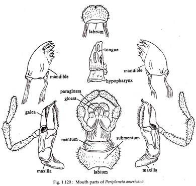
(c) Labium:
Behind the first pair of maxillae are the second pair of maxillae, collectively called the lower lip or labium. It is divisible into a proximal part called sub- mentum and a distal part, mentum.
The sub- mentum articulates with the head just near the articulation of maxillae. The free end of mentum carries many jointed labial palps, outer small paraglossae and inner, much smaller glossae, the latter having curved claw at the tip. It helps to push the food into the mouth cavity.
(d) Hypo-pharynx:
The hypo-pharynx is a fleshy, sensory structure also termed as tongue or lingua. It is centrally placed bounded dorsally by mandibles, ventrally by the labium and laterally by maxillae. A membrane from the inner border of the labium is continuous with its ventral side. The salivary duct opens in the middle of the ventral side and near its base.
(e) Labium:
Labrum or upper lip is a broad oblong plate, movably hinged with the distal end of clypeus. It overhangs the mouth aperture and lies over the mandibles. Its dorsal side is hard but ventral side is soft and is known as epipharynx. It helps in seizing the food.
B. Thorax:
A short neck or cervicum connects the head with the thorax. The thorax is made up of three segments— prothorax, mesothorax and metathorax. Neck is the forward extension of the prothorax. A large sclerite covers the prothorax dorsally. It extends anteriorly to cover the head and posteriorly to shield the mesothorax. This sclerite also extends laterally.
The central part of this sclerite is lighter in colour than its periphery. A slender line runs along the middle and bifurcates posteriorly. The mesothorax is covered by a round sclerite having a central triangular marking called the scutellum. A transverse line above the pointed apex of the scutellum divides the sclerite into an anterior and a posterior part.
The anterior part, called pre-scutum or preallar sclerite, looks like an independent sclerite but actually it is a part of the mesothoracic sclerite. Posterior to the pre-scutum lies the scutum, which on each side bears one anterior and one posterior tergal processes for the articulation of the fore wing.
The exoskeleton of metathorax resembles that of mesothorax, but in metathorax the pre-scutum is not well marked. The anterior and posterior tergal processes are present on the lateral side of the scutum for the articulation of the hind wing.
(a) Legs:
Each thoracic segment carries a pair of jointed, walking legs. All the three pairs of legs are of similar shape and structure. The first pair of legs are the smallest while the third pair are the largest. Each leg (Fig. 1.121) consists of the following parts.
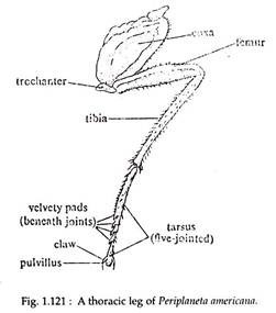
(i) Coxa:
Coxa is the proximal part of the leg and is stout, broad and flat. With the help of this, the leg is articulated with the thorax.
(ii) Trochanter:
It is a small part which serves as a joint between the coxa and the next part, the femur.
(iii) Femur:
It is the long, stout and flat portion, which is the strongest part of the leg. Its outer border is spiny.
(iv) Tibia:
The femur is connected to a slender and long portion called tibia. It has numerous spiny projections on its lateral sides called tibial spurs.
(v) Tarsus:
This is the distal part of the leg following tibia. It is five jointed and each part is called a podomere. Beneath the joints there are soft pads called plantulae. The terminal podomere is known as pretarsus, which bears two claws and a thin hairy pad called arolium or pulvillus. All the legs are used for running, walking and climbing.
(b) Wings:
An adult cockroach has a pair of wings on the dorsal side of mesothorax and metathorax. Each wing is composed of double layers of chitin enclosing tubular tracheae. The chitin thickens around the tracheae to form nervures or veins that strengthen the wings. The wing veins may form a basis of classification as they have different patterns in different insects.
The forewings or mesothoracic wings are darker, brownish, opaque and leathery in texture. They serve to protect the hind wings during rest and are not useful for flying. They are hence known as wing cover or tegmina or elytra. The hind or metathoracic wings are thin, membranous, transparent and broad and are used in flight. At the time of rest, they are kept folded like a fan.
C. Abdomen:
The abdomen is broad, flattened dorsoveritrally and is the longest tagma of the body. It is divisible into ten segments. Last few segments are short and closely set. The last part of the segment in both the sexes is modified to take part in the formation of an area where reproductive ducts open and the area is provided with certain genital appendages (Fig. 1.122).
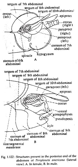
The last part of the abdomen in a female cockroach (Fig. 1.122A) comprises of the following parts.
(a) Hypogynum:
It is formed by the close apposition of the sternites of both the sides of seventh abdominal segment towards the posterior direction. In between these two sternites, a fleshy lobe is placed with one small anterior and a large posterior fold. Dorsally the two folds bear a structure called paraprocts, within which opens the anus.
(b) Epiprocts:
The tergite belonging to the tenth abdominal segment extends posteriorly as incompletely bifurcated flexible projection, carrying three gonapophyses or ovipositors having sclerites called valvifers.
(c) Cerci:
These paired structures are borne by the lateral side of the paraproct and articulate distally with the anterior end of the epiproct and the tergites of the tenth abdominal segment. In addition to the above mentioned structures, the eighth abdominal segment bears three sclerites.
They are two laterals or basisternite and a single median. The spermathecal apertures of female open within the posterior projection of median sclerite. Vulva or female gonopore is present on the ventral side of the two basisternites.
The male cockroach also exhibits specialisation of the posterior end of the abdomen (Fig. 1.122B). The sternum of the ninth abdominal segment is drawn anteriorly up to the seventh abdominal segment and bears cerci, epiprocts, styles and gonapophyses. Cerci and epiprocts are similar in structure to that of the females. The stylets are paired structures and are present only in the males.
They are rudimentary, undivided and present on the ninth segment. Three gonapophyses, two lateral and one ventral are placed within a chamber formed dorsally by paraprocts and ventrally by the sternum of ninth abdominal segment. In between the lobes of the gonapophyses opens the duct from the conglobate gland. The left gonapophysis bears a slender pseudopenis.
4. Integumentary System of Periplaneta:
The integument of cockroach consists of basement membrane, epidermis and cuticle. The basement membrane is the innermost, extremely thin, stellate cell layer. Above it is a monolayer of closely packed hexagonal cells called epidermis and outer to it is the non-cellular layer, the cuticle.
Cuticle is secreted by the epidermis and it externally covers the entire body of the cockroach. It also lines the inner wall of fore gut, hind gut, trachea and genital atrium. Numerous fine tubules originating from the lower epidermal cells traverse the cuticle. Further, the stiff, immovable bristles or spines covering the legs are the outgrowth of cuticle.
The cuticle is divisible into an outer thick epicuticle and an inner thick precuticle. The innermost part of the epicuticle is formed by a substance called arthroidin. Its outer part is a thin polymer layer that is coated externally by a substance called amphion, formed by combination of wax and cement. The amphion makes the cuticle impermeable to water.
Glands:
The integumentary glands of cockroach are cervical glands and abdominal glands. The cervical glands are present within the membrane which covers the neck and its product is called ‘periplanetin’.
The abdominal glands include dorsal abdominal gland in between the terga of 5th and 6th abdominal segments and ventral abdominal gland in between the sterna of 6th and 7th abdominal segments. These abdominal glands produce a substance having pungent smell which is used for defence.
5. Muscular System of Periplaneta:
The muscles of cockroach are classified into two broad groups—skeletal muscles and visceral muscles. There are nearly 370 pairs of skeletal muscles of which 51 are present in the head. Skeletal muscles are present in the mouth parts, thoracic legs, wings and genital appendages. The wing muscles of male are opaque and pink, while those in females are hyaline and white.
The visceral muscles contribute to the lining of gut and heart. The wall of the gut contains an outer coat of circular muscles and an inner coat of longitudinal muscles. The longitudinal muscles are narrow in the posterior region of crop and in the mid gut. While it is strongly developed in the anterior region of crop, colon and rectum.
The heart muscles comprise of fan-shaped alary muscles and a thin circular layer with distinct nuclei. The histology of heart muscles exhibit the presence of intercalated discs in between the muscle cells, a plasma lemma having intimate connection with endoplasmic reticulum and complex mitochondria between the myofilaments.
6. Body Cavity of Periplaneta:
The body cavity in the form of coelom is present only in the embryonic condition. In adult cockroach, the body cavity is formed by the fusion of embryonic blastocoel with the embryonic coelomic space and is called mixocoel. The wall of the embryonic coelom is used in the formation of different organs. The mixocoel in cockroach is obliterated by a loose tissue called fat bodies.
The rest of the space is occupied by the digestive, excretory and reproductive organs. Blood flows through the mixocoel and for this reason the mixocoel is also called haemocoel and the circulating fluid is called haemolymph.
The cells in the fat bodies are of two types. One type is bi-nucleated and the other type is with an elongated nuclei. These cells act as store-house of reserve food which remain in the form of glycogen and are used during starvation.
7. Locomotion of Periplaneta:
The locomotory organs of cockroach are the legs and wings. All the three pair of legs are used in walking, running or climbing.
8. Digestive System of Periplaneta:
Cockroaches are omnivorous. Its digestive system comprises of alimentary canal and digestive glands. Extracellular digestion is the characteristic of cockroaches.
9. Respiratory System of Periplaneta:
Respiration in cockroach is aerial.
10. Circulatory System of Periplaneta:
Cockroach possesses an open circulatory system which is poorly developed. The circulating fluid is called blood or haemolymph. It consists of a colourless fluid called plasma, suspended in which are many haemocytes (9 millions in a 24 hrs. old adult).
11. Excretory System of Periplaneta:
Excretion in cockroach is chiefly conducted by the malpighian tubules. Other accessory excretory structures of cockroach are nephrocyte or pericardial cells, rectal glands, fat cells and some amoeboid cells.
12. Nervous System of Periplaneta:
The nervous system of cockroach comprises of:
(a) Central nervous system comprising of a brain or supraoesophageal ganglion, sub-oesophageal ganglion, circumoesophageal connectives and the ventral nerve cord.
(b) Peripheral nervous system made up of nerves given out from the ganglia of the central nervous system.
(c) Sympathetic nervous system is formed by a ganglionated sympathetic nerve.
(d) Sense organs are represented by compound eyes, ocellus, cuticular hair, chordotonal organs etc.
13. Endocrine System of Periplaneta:
Cockroaches contain certain endocrine organs such as:
(i) Corpora Cardiaca,
(ii) Corpora Allata,
(iii) Prothoracic Gland and
(iv) Cervical Glands.
These glands work together along with five groups of specialised cells in the brain. Of these five groups, three are placed anteriorly and they form the first nerve to the corpora cardiaca. The remaining two groups are present at the posterior end and they send the second nerve to the corpora cardiaca.
(i) Corpora Cardiaca:
This is a paired small, elongated and irregular gland, whose anterior end encloses the aorta. Its dorsal side bears a transverse commissure which extends posteriorly as a process to give rise to the aortic nerves. Ventrally, the corpora cardiaca are connected with a rudimentary ganglion and are drawn posteriorly to connect the corpora allata.
Each corpora cardiacum receives three nerves of which two are from the neurosecretory regions of the brain and one from unknown origin. Electron microscopic studies revealed the profuse presence of endoplasmic reticulum and secretory granules of 600 mm diameter, in the cells of corpora cardiaca.
The secretion of corpora cardiaca affects the contractility of muscles, lining the gut, the malpighian tubules and heart. It is believed that the products released by the corpora cardiaca increase the effects of prpthoracic glands.
(ii) Corpora Allata:
Posterior to the corpora cardiaca lies a pair of small glands, the corpora allata. It is more or less oval in shape and a transverse commissure connects it with the oesophageal nerve. It has nerve connections with sub-oesophageal ganglion, corpora cardiaca and prothoracic glands.
Histologically, the cells of corpora allata shows the profuse presence of mitochondria but the secretory granules are present in limited areas. The secretion of corpora allata maintains the juvenile features in the larval stage, helps in the oocyte formation in adult females and influences the secretion from secondary sexual organs in both the sexes.
(iii) Prothoracic Glands:
At the anterior end of the prothoracic ganglion, lies a pair of rope-like prothoracic glands. A tracheal branch remains associated with the gland. Histologically, prothoracic gland shows the presence of a central part having 6-8 muscle fibres, which remain enclosed by an outer glandular covering of 4-12 cells deep. The secretory granules are of various sizes.
The secretions of prothoracic gland facilitate moulting and after the last moult it degenerates. It has been experimentally demonstrated that the implantation of pairs of prothoracic glands in an adult induces it to moult and results into the formation of a giant cockroach.
(iv) Cervical Glands:
In the neck region near the posterior opening of the head is present a pair of small oral, cervical glands, that are richly supplied with trachea. The outer part of this gland is composed of large glandular cells and the inner part is made up of small cells with unusually large nucleus.
The central part of the gland is occupied by a cavity which traverses within the inner wall. The endocrine nature of this gland has not yet been worked out in cockroach, but in other insects it is believed to be responsible for producing a hormone to induce moulting. The product which is secreted is called periplanetin.
14. Reproductive System of Periplaneta:
Sexual dimorphism is well marked in cockroaches and the two sexes can be easily identified on the basis of their morphological features. Cockroach undergoes sexual reproduction.