
The below mentioned article provides an overview on Ascaris:- 1. Introduction to Ascaris 2. Habit and Habitat of Ascaris 3. Structures 4. Body Wall 5. Body Cavity 6. Locomotion 7. Digestive System 8. Respiratory System 9. Excretory System 10. Nervous System 11. Reproductive System 12. Life History 13. Pathogenicity 14. Therapeutic Treatment.
Contents:
- Introduction to Ascaris
- Habit and Habitat of Ascaris
- Structures of Ascaris
- Body Wall of Ascaris
- Body Cavity of Ascaris
- Locomotion of Ascaris
- Digestive System of Ascaris
- Respiratory System of Ascaris
- Excretory System of Ascaris
- Nervous System of Ascaris
- Reproductive System of Ascaris
- Life History of Ascaris
- Pathogenicity of Ascaris
- Therapeutic Treatment of Ascaris
1. Introduction to Ascaris:
These are stout worms of fairly large size. Males measure about 15-25 cm in length and diameter of 3-4 mm, while females have 25-40 cm length and maximum 5 mm diameter. Their mouth is provided with three conspicuous lips — one dorsal and two ventral. Species of genus Ascaris inhabit the intestine of various mammals.
ADVERTISEMENTS:
The most common and best known member is Ascaris lumbricoides, the large intestinal roundworm of man causing ascariasis. This worm was observed and reported by many ancient people but details about its life cycle were known only after 1916.
Etymology:
Greek: askaris, pinworm
The species name lumbricoides (Latin: Lumbricus, earthworm; and Greek: eidos, resemblance) was given because of its similarity to the earthworm.
2. Habit and Habitat of Ascaris:
ADVERTISEMENTS:
The adult worm is an endoparasite residing in the small intestine of man. The parasite, although normally an inhabitant of the jejunum, may wander up and down the intestinal tract and may even penetrate into other neighbouring channels. It has a cosmopolitan distribution, but are most frequent in tropical and subtropical countries. It is common particularly among children.
3. Structures of Ascaris:
The adult worm is light brown or pinkish in colour when alive, but it gradually changes to white, when present outside the intestine. Its body is cylindrical in shape tapering at both ends. The anterior end is thinner than the posterior end (Fig. 1.64A & B).
There are four longitudinal bands or lines with a number of fine transverse striations all over the body. Of these four streaks, one is dorsal, one ventral and two lateral in position. The dorsal and ventral streaks are narrow and pure white in living condition, while the lateral ones are thick and light brown in colour.
The mouth aperture opens at the anterior or terminal end and is bounded by three lips, one dorsal and two ventral (Fig. 1.64C).
ADVERTISEMENTS:
The lips are semi-circular in shape, being finely toothed and having dentigerous ridges. The mouth is marked off by a sharp constriction from the rest of the body.
A little on the ventral side of the posterior end is situated the anus, that opens directly in the females as a transverse slit (Fig. 1.64H), whereas in the male it opens with the ejaculatory duct into the cloaca. A little down the anterior tip and on the ventral surface is situated an excretory pore.

Ascaris exhibits sexual dimorphism. Female worms are larger and stouter than the males. Females can be distinguished by the presence of separate and independent genital aperture situated on the ventral surface at about one-third of the body length from the anterior.
There is no separate male genital aperture. Sexes can also be distinguished externally by the shape of the post-anal portion of the body. The post-anal portion in the male is sharply curved downwards while in the female it is nearly straight. At the curved portion of the males a pair of needlelike chitinoid bodies project from the cloacal aperture, called penial setae (Fig. 1.64F & G).
4. Body Wall of Ascaris:
The body wall of Ascaris is made up of an outer layer of cuticle which is thin, transparent, delicate and wrinkled transversely (Fig. 1.65). The cuticle of Ascaris produces enzyme inhibitors that protect it from the host’s digestive enzymes. Beneath the cuticle is the ectoderm which forms a syncytial protoplasmic layer. The ectoderm is responsible for secreting the cuticle.

Below the ectoderm is a single layer of longitudinal muscle. The individual cells of the longitudinal muscles are spindle-shaped, straightened longitudinally and bears at the middle a bladder-like mass of protoplasm containing the nucleus which faces the side of the body (Fig. 1.66). This peculiar appearance of the longitudinal muscles is due to the differentiation of the cell into a contractile and a nuclear part.
The longitudinal muscle layer is not continuous and is arranged into four longitudinal bands, two dorsolateral and two ventro-lateral (Fig. 1.65). This is due to the fact that the ectoderm projects inwards at the dorsal, ventral and two lateral sides between the muscles to give rise to the streaks on the outer surface of the body.

5. Body Cavity of Ascaris:
The body cavity is not a true coelom as it is not lined by cell layers derived from the mesoderm. It, however, develops from blastocoel (i.e. between mesoderm and endoderm of embryo) and is referred to as pseudocoel.
The body cavity is linked externally by fibrous processes of the longitudinal muscle cells and internally by cuticle encasing the intestine. There are five giant stellate mesenchymal cells in the pseudocoel of Ascaris, called pseudo-coelomocytes, occupying fixed positions. Pseudocoel is filled with an odorous protein rich fluid, known as perienteric or pseudocoelomic fluid.
6. Locomotion of Ascaris:
To counteract the peristaltic activity of the host’s intestine, the dorsolateral and ventro-lateral muscles in the anterior end of Ascaris, contracts alternately to perform undulating movements. However, ability to perform change in body length is restricted due to absence of circular muscles in the body wall.
7. Digestive System of Ascaris:
The alimentary tract is a straight tube which runs along the entire length of the body (Fig. 1.67). Mouth is anterior and terminal in position and is guarded by three semicircular lips that are finely toothed. Behind the mouth is the buccal cavity, which leads into a club-shaped pharynx. The pharynx is dilated and its wall is muscular, which helps it to draw food from the intestinal content of the host.
Pharynx then leads to the intestine, whose posterior part narrows down to form the rectum. The rectum opens to the outside through the anus, which lies about 2 mm from the tail end. The entire alimentary canal is made up of a single epithelial layer covered internally and externally by cuticle derived from the ectoderm. Digestive glands are totally absent as Ascaris feeds upon the digested food of the host.

8. Respiratory System of Ascaris:
Like most other endoparasites, Ascaris respires anaerobically, as the oxygen content in the host’s intestine is generally poor. According to Hyman, the small amount of haemoglobin present in the pseudocoelomic fluid and body wall, serves to transport oxygen.
9. Excretory System of Ascaris:
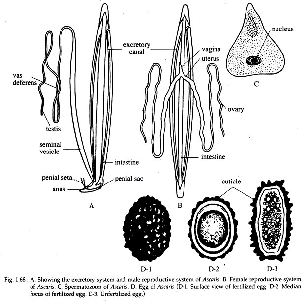
The excretory system of Ascaris consists of two longitudinal excretory canals, one through each lateral streaks (Fig. 1.68A). The two canals are connected anteriorly, below pharynx, by a transverse canalicular network. Extending from this is a short terminal excretory duct that opens to the outside through the single excretory pore situated on the ventral side.
Each longitudinal excretory canal extends posteriorly along the entire body length through a lateral epidermal chord and remains closed at its terminal end. Four to six big tufts of cells with ramifying protoplasmic processes remain in close contact with the canals.
It is believed that these cells collect, store and pass on the waste matter in dissolved state to the excretory canals. The whole system is neither a ciliated one nor there is any flame cells. It is presumed that the canal is an intracellular tubular extension of a single enormously elongated cell.
10. Nervous System of Ascaris:
The nervous system of Ascaris comprises of a nerve ring or circumenteric ring around the pharynx (Fig. 1.69). The ring is swollen at the ventral side and is ganglion like. The ring gives off six nerves each to the anterior and posterior sides.
Two of the posterior nerves are of considerable thickness and run along the dorsal and ventral lines up to the posterior end of the body. The dorsal and ventral longitudinal nerves are connected with each other by transverse commissures. The posterior tip of the ventral nerve cord is swollen and forms a ganglion just in front of the anus. The sense organs are the sensory papillae situated as small elevations on the lips.
11. Reproductive System of Ascaris:
Sexes are separate and sexual dimorphism is well marked (Fig. 1.64). Gonads are typically long, coiled and tubular. They are attached at the genital pore in females and cloaca in males. The male reproductive organs consist of a single coiled, thread-like tubule (Fig. 1.68A) occupying some portion of the body cavity and about eight times the length of the body.
This structure is differentiated at the anterior region into the testis, the middle region as the vas deferens and the posterior region as the seminal vesicle. The demarcations of these parts are very poor. The seminal vesicle continues as the ejaculatory duct and opens into the anus.
At the opening of this structure there is a pair of chitinoid spicules called penial setae, each of which is provided with a muscular penial sac. In case of male Ascaris, the anus is used for the elimination of faeces as well as of sperms and therefore, is called cloaca.
The sperms of Ascaris (Fig. 1.68C) are very peculiar as they show amoeboid movement inside the body of the female. A matured sperm is cone-shaped, having a broad base and an apex. The apex contains the acrosome and the broad base contains the nucleus and mitochondria. The sperms remain non-motile in the male gonoduct.
The female genital organs are a much coiled structure (Fig. 1.68B) present in the middle and posterior third of the body and its length is several times more than the entire length of the worm. If comprises of a pair of much coiled thread-like ovaries which pass into an uterus.
The two uteri unite to form a single, conical and muscular vagina, which opens to the outside by the female genital aperture situated on the ventral surface at about one-third the body length from the head. The fertilized and unfertilized eggs vary in character (Fig. 1.68D).
The fertilized egg is round or oval in shape, measuring 60 to 70 µm in length and 40 to 50 µm in breadth. It is always bile-stained and brownish in colour. It contains a very large conspicuous, un-segmented ovum, the nucleus of which is concealed by a large amount of coarse yolk granules. It is surrounded by a thick smooth translucent shell with an outer albuminous coat having a rippling surface.
The unfertilized egg on the other hand is narrower, longer and more elliptical in shape. It has a thinner shell with an irregular coating of albumen and contains an atrophied ovum. The eggs of Ascaris are highly resistant to desiccation and disinfectants and may remain viable in soil for 10 years.

12. Life History of Ascaris:
Ascaris passes its life cycle (Fig. 1.70) in one host and no intermediate host is required. Man is the only known definitive host. Eggs are produced in large numbers (20,000 a day) and are fertilized in the upper part of the uterus. This takes place when the parasite remains in the intestine of the host. The egg after fertilization gets enclosed in a chitinous egg shell.

The egg in this condition is passed to the outside along with the stool of the host. At this stage they are not infective to man. The development of the zygote to larva takes place outside the body of human host and it takes a fairly long time (about 10-40 days), depending upon the atmospheric temperature and humidity.
The egg containing the coiled-up embryo is infective to man. The embryo does not hatch in the open but remains quiescent within the egg shell until it reaches a new host.
Infection to the new host is direct. Infection results when contaminated food or drinks are ingested by man. The coiled-up embryonated eggs pass down to the duodenum where the digestive juices weaken the egg shell and stimulate the enclosed larvae into activity.
The larvae emerge through a rent in the egg shell produced as a result of splitting and not by digestion of the eggshell. It takes about two hours to hatch. These larvae measure about 0.2 to 0.3 mm in length and 13 to 15 µm in breadth.
The larvae undergo moulting before hatching. The newly hatched larvae burrow their Way through the mucous membrane of the small intestine and enter the lymphatic ducts or veinules. From the lymphatic ducts it is carried to the mesenteries, lymph nodes and then to the portal vein from where the larva comes to the liver. Here, they live for a period of 3 to 4 days.
Then, after passing through interlobular veins, central veins, sub-lobular veins and hepatic veins, are drained into the inferior vena cava which opens into the right atrium of the heart. From here they are carried to the lungs, where the larvae moult twice and grow much bigger in length (from 0.2 mm to 2.0 mm). Breaking through the capillary wall they reach the lung alveoli and crawl up the bronchi and trachea.
They are then propelled into the larynx and then to the pharynx where they are once more swallowed. It takes about 10 to 15 days for such migration from the lungs to the pharynx. The larvae, then pass down the oesophagus to the stomach and upper intestine, where the fourth moulting takes place. They while staying in the lungs acquire a resistance to the gastric juice.
The larvae having returned to their normal abode become much bigger, about 10 times their original size and measuring about 2 to 3 mm in length. In about 6 to 10 weeks-time, they become sexually mature. The worm starts producing eggs within two months from the time of infection. The life-span of the adult worm in the human host is rather short, average being less than a year.
At the time of massive infection, the larvae may reach the general circulation and are carried to the various organs and tissues. They may then happen to lodge in such aberrant sites as kidney, brain, spinal cord and other organs. They are unable to grow to maturity and most of them are destroyed. They may even be secreted in the urine at the time of heavy infection.
13. Pathogenicity of Ascaris:
Mode of infection:
Infection may take place either by swallowing fully developed Ascaris eggs encased in shells, with raw vegetables or by drinking contaminated water. It may also be conveyed directly into the mouth through dirty fingers. Incidence of Ascariasis is greater in children than in adults.
Incubation period:
In man it takes 60 to 75 days from the time of exposure of infection, for the mature female to lay eggs and that is the period when symptoms are manifested.
Pathogenesis:
The adult worm produces the following pathogenic effects:
(i) Spoliative action:
By robbing the host of its nutrition.
(ii) Toxic action:
The body fluid of Ascaris is toxic and causes anaphylactic reaction.
(iii) Mechanical effect:
By causing obstruction of the tubular passages, such as appendix, bile duct, pancreatic duct, bronchus and perforating through ulcers in the alimentary canal.
14. Therapeutic Treatment of Ascaris:
For a long time santonin was the classical remedy of ascariasis but recently oil of chenopodium and hexylresorcinol have been widely used. Seeds of Butea and dried fruits of Embelia have also been used by grinding it into a paste and mixing it with sugar and water.