The below mentioned article provides an overview on Asterias:- 1. Introduction to Asterias 2. Habit and Habitat of Asterias 3. Structure 4. Integumentary System 5. Skeleton 6. Section of Arm 7. Body Cavity 8. Digestive System 9. Respiratory System 10. Water Vascular System 11. Locomotion 12. Haemal System 13. Axial Complex and Other Details.
Contents:
- Introduction to Asterias
- Habit and Habitat of Asterias
- Structure of Asterias
- Integumentary System of Asterias
- Skeleton of Asterias
- Section of Arm in Asterias
- Body Cavity of Asterias
- Digestive System of Asterias
- Respiratory System of Asterias
- Water Vascular System of Asterias
- Locomotion in Asterias
- Haemal System of Asterias
- Axial Complex of Asterias
- Excretory System of Asterias
- Nervous System of Asterias
- Oral or Ectoneural System of Asterias
- Sense organs in Asterias
- Reproductive System of Asterias
1. Introduction to Asterias:
The members of the genus Asterias are generally called the ”star fishes” or ”Sea-stars”. They are star shaped, free-moving echinoderms distributed in all the oceans and at all depths. Sea stars are commonly red, orange, blue, purple or green in colour or exhibit combination of colours.
ADVERTISEMENTS:
In the Andaman area purple and pink coloured star fishes can be seen. There are about 150 species under the genus Asterias of which some important ones are A. rubens, A. gibbosa, A. vulgaris, A. forbesi, A. amurensis, A. panceri etc.
Etymology:
Greek: Aster, star
2. Habit and Habitat of Asterias:
Asterias is a marine and widely distributed member of echinoderm. All the species under this genus are benthic animals, as they inhabit the bottom of the sea. They are quite abundant on various types of sea-bottom but prefers a rugged and rockbound coast, where they can hide very easily and lead a sluggish life. They also live in deep water.
ADVERTISEMENTS:
They are carnivorous and predacious animals, preferring mainly bivalves as food. Majority of the forms are photonegative and prefer to live in shaded areas. A few exceptions are the Asterias rubens, A. gibbosa, A. panceri, where positive response to light is observed.
3. Structure of Asterias:
The body of Asterias is flattened in the oral-aboral axis and has a five-pointed star- shaped body (Fig. 1.131). The body consists of a central disc and five symmetrically placed arms (or rays). The arms are conspicuously broad at their bases and they gradually taper towards the tip.
The lengths of the arms are two or three times the diameter of the disc. The body exhibits radial symmetry and has two surfaces. The upper, aboral or abactinal surface is convex and much darker, while the lower, oral or actinal side is flat and less pigmented.
The imaginary lines dividing the central disc and the arms are called the radii and the intervening regions between the radii are called inter-radii. Body is covered by a hard and tough covering containing numerous calcareous ossicles.
ADVERTISEMENTS:
At the middle of the central disc, on the oral surface, a five-rayed aperture called mouth or actinosome (Fig. 1.131B) is present. This opening is surrounded by a membranous peristome and it also possesses five groups of oral papillae. Five narrow ambulacral grooves, one on each arm, run orally from the five-rayed aperture to the tip of the arms.
The edges of these grooves are provided with two or three rows of movable calcareous ambulacral spines. Besides the above, rows of immovable stout spines are also present. The aboral side contains numerous irregular rows of short and stout spines supported by ossicles.
The oral and aboral surfaces of the body are separated by a row of prominent spines. Many dermal pores are situated in the spaces between the ossicles on the aboral side.

At the centre of the aboral side, a minute aperture called anus, is situated. On the same aboral side, between the bases of two of the five arms (bivium), is situated a structure called madreporite (Fig. 1.131A). The madreporite is a flat disc-like body with radiating grooves. The presence of more than one madreporite, in some species, is possibly due to increase of the number of arms of more than the usual five.
Scattered all over the body surface are many microscopic bodies called pedicellariae (Fig. 1.132A). Each pedicellariae consists of a flexible stalk and three calcareous pieces—one basilar and two jaws or valves. The two jaws are attached to the basilar piece.

This particular variety of pedicellariae is called the pedunculate, of which there are two types (Fig. 1.132B) in Asterias.
These are:
ADVERTISEMENTS:
(i) Straight Pedicellariae, where the two jaws remain more or less straight on the basilar piece and
(ii) Crossed Pedicellariae, where the basal portions of the jaws are curved and cross each other.
The pedicellariae are modified spines and are regarded as protective organs. The jaws are movable on the basilar piece, operated by two sets of muscles — two pairs of adductor muscles to close them and one pair of abductor muscles to open them.
In natural condition and in well- preserved state two double rows of tube feet or podia are seen in the ambulacral grooves. Each tube foot or podium has a soft tubular body that ends in a sucker. These are locomotory organs and are capable of great extensions.
At the tip of the ambulacral groove, sense organs are placed. A minute red spot, called the eye, is present in the extremity of each ambulacral groove on the oral side. The eye is composed of several ocelli. Just above the eye lies a median unpaired terminal tentacle. This tentacle resembles a tube foot but lacks the sucker and is regarded as olfactory organ.
4. Integumentary System of Asterias:
The body wall of Asterias is covered externally by cuticle, which is differentiated into an outer thick homogeneous layer and a very delicate inner layer. Beneath the cuticle is situated the epidermis (Fig. 1.133).
The shape and structure of the cells composing the epidermis vary greatly at different regions of the body. Usually these cells are of ciliated columnar types. Besides the above several other cells are also present, such as neurosensory cells, pigment cells and glandular cells.
The gland cells are of two types:
(i) Goblet or mucus gland cells that are filled with finely granular substance, providing the body with a protective mucous coating and
(ii) Muriform gland cells that are filled with coarse granules. At the base of the epidermis lies a network of nerve fibrils.
Below the nerve fibrils lie the dermis which is comparatively thicker. It is mesodermal in origin and is composed of fibrillar connective tissue. The epidermis and dermis are separated by a delicate basement membrane.
The dermis contains the endo-skeletal ossicles and also possesses a system of canalicular haemal spaces. At the inner end of the dermis lies the muscular layer, which is divided into an outer circular muscle layer and an inner longitudinal muscle layer. The innermost layer of the body wall is lined by the coelomic epithelium.

5. Skeleton of Asterias:
The body of Asterias is quite rigid as well as capable of considerable degree of binding and twisting. The rigidity of the body is due to the presence of definite skeleton, made of supporting skeleton and superficial skeleton.
The supporting skeleton comprises of the main deeper skeletal elements (ossicles) embedded in the dermis and the superficial skeleton are in the form of spines, warts and tubercles which are borne on the deeper skeleton. The ossicles are of various shapes and bounded together by connective tissue.
The ossicles have distinct pattern of arrangement. The mouth is surrounded by five plate-like oral ossicles. Each ambulacral groove is supported by double rows of rod-like ambulacral ossicles. These ossicles are articulated with the adambulacral ossicles at their aboral ends like an inverted ‘V’.
The apex of the ‘A’ projects into the coelom and forms a prominent ambulacral ridge. The ambulacral ossicles are movably articulated so as to allow the closure and opening of the ambulacral groove. A row of lateral adambulacral ossicles meet the ambulacral ossicles at their outer ends. The adambulacral ossicles bear moveable spines.
In some forms of Asterias, one or two rows of additional ossicles are present, called supra-marginal and infra-marginal ossicles depending on their positions. The .marginal ossicles form the dorsolateral sides of the arms and usually remain covered by small ossicles. A series of carinal ossicles form the mid-dorsal skeleton of the arms. Between the carinal and marginal ossicles there may be a number of elongated dorsolateral ossicles.
The arrangement of skeletal elements on the aboral side is indistinct in adults, but distinct in a freshly metamorphosed Asterias. At the middle of the aboral side a central plate is present which bears the anus. This central plate is surrounded by five inter-radial plates. One such plate incorporates the madreporite.
The inter-radial plates are surrounded by radially placed terminal plates. In star fishes, where the number of arms exceed five, the inter-radial and terminal plates increase with the number of arms. As the arms grow in length the terminal plates are also shifted to the tip and additional row of plates develop behind it. These plates which develop behind the terminals constitute the carinals of the mid-dorsal line of the arms.
6. Section of Arm in Asterias:

Any one of the arms when sectioned shows the following structures (Fig. 1.134):
i. The dorsal wall of the arm presents the appearance of an arch and the ventral part is shaped like an inverted ‘V’, the ambulacral furrow. The aboral side is thicker than the oral side.
ii. The body wall consists of epidermis, dermis, muscular layer and coelomic epithelium. The epidermis consists of ciliated columnar cells, neurosensory cells, pigment cells and two types of gland cells. On the upper surface of the epidermis lies the cuticle and beneath the epidermis is the basement membrane.
Dermis is mesodermal in origin, fibrillar in nature and lies beneath the basement membrane. The dermis is thicker than the epidermis and contains haemal spaces and endoskeleton in the front of ossicles. The lower layer of the dermis contains the muscle layer, which is differentiated into an outer circular layer and an inner longitudinal layer. The coelomic epithelium forms the inner lining of the body wall.
iii. The surface of the body bears spines, tubercles, pedicellariae and warts which are borne on the deeper skeletal elements.
iv. The deeper skeleton consists of ossicles of various shapes. The ambulacral groove is supported by two rod-like ambulacral ossicles, articulated at their aboral ends with the upper transverse ambulacral muscles and appearing like an inverted ‘V’.
Two adambulacral ossicles are present, one on either lateral side of each ambulacral ossicle. The ambulacral ossicles bear movable spines. The upper dorsomedian side of the arm contains carinal ossicle. Between the carinal and adambulacral ossicles there exist supra- and infra-marginal ossicles which form the lateral walls of the arm.
v. Projecting between the dermal ossicles there are many hollow outgrowth of the body called respiratory papulae that are quite abundant on the aboral surface.
vi. The spacious cavity enclosed by the body wall is called coelom and scattered in it are the coelomocytes.
vii. Along the ambulacral groove are present double rows of tube feet. Each tube foot has a bladder-like ampulla ending in a sucker. The ampullae are situated in the cavity of the arm.
viii. Suspended in the cavity of the arm are two pyloric caeca by means of two mesenteries, from the aboral body wall.
ix. Two gonads are located in the cavity of the arm between the pyloric caeca and the ampullae.
x. Towards the oral side of the arm are present the radial canal and lateral or podial canals of the water vascular system.
xi. The oral side also contains the radial nerves, Lange’s nerve, hypo-neural sinus with its dividing septum that contains radial haemal strand.
7. Body Cavity of Asterias:
The body cavity of Asterias is a true coelom, which is distinct and reaches to every part of the body. The coelom is enterocoelic in origin. There is a large perivisceral coelom which extends into the central disc and surrounds the alimentary canal and the gonads. There are many small coelomic cavities like water vascular system, axial sinus, perihaemal cavities and genital sinuses.
The inner surface of the perivisceral coelom is lined by ciliated epithelium which is differentiated into two separate layers. The parietal layer covers the inner surface of the body wall and the visceral layer forms an investment for the different organs contained in the body.
The coelom is filled with a coelomic fluid, which is chemically similar to sea water in composition and contains amoeoid coelomocytes. These are of two types— amoebocytes with ordinary slender pseudo- pods and amoebocytes with petaloid pseudopods. Both are regarded to be the two phases of the same cell. The coelomocytes are highly phagocytic and are supposed to be excretory in function. The coelomic fluid of Asterias contains potassium in higher concentration than in sea water
Traces of amino nitrogen are reported to be present in asteroids. A small amount of reducing sugar is present in peri-visceral fluid of Asterias forbesi. The coelomic fluid also contains excretory products like urea and ammonia.
8. Digestive System of Asterias:
Digestive system of Asterias consists of a very short straight alimentary canal and digestive glands.
9. Respiratory System of Asterias:
Respiration in Asterias is carried out by numerous delicate dermal papulae. The papulae are simple, contractile, transparent and hollow outgrowths of the body wall (Fig. 1.132C) that project between dermal ossicles. These are distributed abundantly on the aboral side of the body.
The histological structure is same as that of the body wall excepting that, the dermis is thinner and dermal skeleton is totally absent. The lumen of each papula is continuous with the coelom and is lined by ciliated coelomic epithelium.
Through these respiratory papulae exchange of oxygen and carbon dioxide takes place by diffusion. In addition to the papulae, the water vascular system also helps in respiration. The tube feet of Asterias rubens are responsible for approximately 10% of total oxygen uptake.
10. Water Vascular System of Asterias:
The water vascular system is the most characteristic system in echinoderms and plays a number of vital functions. In Asterias it starts with the madreporite (Fig. 1.135A) and gives off a system of vessels traversing the body. The madreporite is a round calcareous plate (Fig. 1.135B) and has an inter-radial disposition on the aboral surface. The madreporite contains furrows which have numerous pores at the bottom. Each pore leads into a pore canal.
The number of pores and pore canals are about two hundred. The pore canals unite to form collecting canals which open into madreporic ampulla. This ampulla proceeds downwards as an ‘S’- shaped cylindrical madreporic or stone canal. The wall of this canal is supported by a number of calcareous rings, hence the name stone canal (Fig. 1.135D).
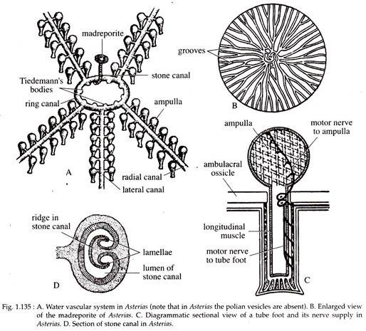
From the wall of the stone canal projects a ridge which bifurcates into two lamellae. The lamella is spirally rolled and occupies a considerable portion of the lumen of the stone canal. Most of the pore canals from the madreporite open into the stone canal, while the rest open into the axial sinus.
The stone canal opens beneath into a considerably wide pentagonal ring canal. In certain starfishes, there occur pear-shaped sacs called polian vesicles. In Asterias, however, polian vesicles are not present. The ring canal in Asterias gives off inter-radially nine small, spherical, glandular structures called Tiedemann’s bodies.
The inter-radius which bears the stone canal has only one Tiedemann’s body. The significance of these bodies is not properly known. However, some workers regard them as lymphatic glands manufacturing amoebocytes of the water vascular fluid.
Some stone canals lie folded in the wall of a wide axial sinus which is a part of the haemal system. The axial sinus communicates with the stone canal aborally and opens to the exterior through some pores of the madreporite. Associated with the stone canal and in-folded in the wall of the axial sinus, lies the axial organ (Fig. 1.135). The actual function of this organ and its relation with the water vascular system is still unknown.
The ring canal gives off five radial canals along the ambulacral grooves of the arms. The radial canals run up to the tip of the arms and end at the lumen of the terminal tentacle. The radial canal gives out small side branches called the lateral or podial canals. Each lateral canal, after reaching the ambulacral pore, divides at right angles into two branches.
One branch is continued as the lumen of the tube foot (Fig. 1.135C), while the other as the cavity of the ampulla. The ampullae are rounded sac like bodies arranged in one or two rows on each side of the ambulacral ridge. Usually one ampulla is present in each tube foot. In Asterias the ampullae are simple and undivided.
The vessels of the water vascular system have muscular wall with an internal epithelial lining. The muscular layer becomes highly developed in the tube feet and ampullae. A muscular valve at the point where the lateral canal joins the lumen of the tube foot, isolates each tube foot and ampulla unit from the rest of the water vascular system, so that the necessary hydrostatic pressure can be built up.
11. Locomotion in Asterias:
Locomotion in Asterias is done by the action of the tube feet. Here, the water vascular system operates on the principle of hydraulic pressure and is indirectly concerned with the movement of the animal.
12. Haemal System of Asterias:
In Asterias, the blood lacunar system as such is absent and its function has been taken up by a special system called haemal system, which consists of channels enclosed by coelomic spaces.
13. Axial Complex of Asterias:
A very well-formed axial complex comprising of a coelomic cavity enclosing the stone canal and axial gland (Fig. 1.136) is present in Asterias. The axial complex remains intimately associated with the inter-brachial septum. The axial sinus is a thin-walled tubular cavity which opens orally into the inner smaller ring of the hypo-neural ring sinus.

It opens aborally into the genital sinus and finally into the ampulla of the stone canal. The axial gland is a long spongy body of brown or purple colour and gives a small terminal process at the aboral end. This process is surrounded by a contractile closed sac named as terminal sac or dorsal sac or madreporic vesicle. This vesicle is situated very close to the ampulla of the stone canal, but has no connection with it.
The axial gland terminates orally in the septum of the hypo-neural ring sinus. The axial gland is variously called as heart, ovoid gland, brown gland, septal organ and dorsal organ. The function of the axial gland and its relation to other systems is still disputed.
It has an interior core of connective tissue traversed by many spaces containing coelomocytes and externally it is covered by a coelomic epithelium. The histological picture reveals its similarity with the haemal system and both are closely related.
14. Excretory System of Asterias:
In Asterias definite excretory organ is lacking. The coelomocytes, however, seem to play the major role in excretion. In addition to other functions, the intestinal caeca also extracts waste products from the coelomic fluid and eliminates it through the alimentary canal.
The nitrogenous products diffuse out from the body into the coelomic fluid and are immediately engulfed by the wandering coelomocytes and pass out of the body through the papulae. The nitrogenous wastes include ammonia, urea and traces of uric acid. The presence of creatin and creatinine in Asterias is very significant as these are not found in any other invertebrates.
15. Nervous System of Asterias:
The nervous system of Asterias is very simple, primitive and diffused type. It includes only nerve net and has no central ganglionic formation. The nervous system consists of oral or ectoneural system, deep or hypo-neural system and coelomic nervous system.
16. Oral or Ectoneural System of Asterias:
This system includes the main part of the nervous system (Fig. 1.137) and is situated below the epidermis. This includes a nerve pentagon situated in the peristomial membrane surrounding the mouth. The nerve pentagon at each radius gives off a radial nerve which runs as a slender thick band throughout the ambulacral groove of the arms.

The radial nerve is situated just below the radial canal as median integumentary thickening. The radial nerve ends as a sensory pad or cushion at the base of the terminal tentacles. The radial nerve gives branches to the tube feet and becomes continuous with the sub-epidermal nerve plexus of the body wall.
The nerve pentagon and the radial nerves consist of nerve fibrils with scattered bipolar and multipolar nerve cells. The radial nerve appears like a thick ‘V’- shaped body below the hypo-neural sinus. It is separated from the hypo-neural sinus by a thin dermis and coelomic epithelium and is covered on its outer side by epidermis.
Deep or Hypo-neural System:
The different nerves composing this system are motor nerves. This nervous system comprises of Lange’s nerves, which is a plate of nervous tissue situated beneath the coelomic epithelium of the hypo-neural sinus and forms a lateral lining on the wall of this sinus. Lange’s nerve extends as five inter-radial nerve thickenings above the main nerve pentagon of the ectoneural system. Each Lange’s nerve innervates the muscles of the corresponding arm.
Coelomic Nervous System:
The sub-epithelial nerve plexus at the outer ends of the ambulacral ossicles form marginal nerve cord on two sides of the arms. The marginal nerve cord gives off lateral motor nerves which proceeds to the aboral side between ambulacral and adambulacral ossicles and forms a plexus beneath the whole of the coelomic lining. It innervates the muscle of the body wall and gonads.
17. Sense organs in Asterias:
The senses are poorly developed and the sense organs in Asterias comprises of the neurosensory cells, terminal tentacles and the eyes.
Neurosensory Cells:
These are present scattered throughout the body surface, but are quite abundant in the suckers of the tube feet, in the ambulacral regions, in the epidermis, at the base of pedicellariae and spines. Each neurosensory cell has a slender fusiform body, the proximal end of which is connected with sub-epithelial nerve plexus and the distal end goes up to the cuticle. These neurosensory cells are tactile in function and act as chemoreceptors.
Terminal Tentacles:
These tentacles are regarded as tactile receptors. They help to survey the environment during locomotion.
Eyes:
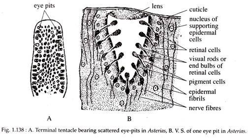
The major sensory organs are the simple, pigmented eye spots situated one at the tip of each arm, on the oral side of the so- called terminal tentacle. The radial nerve cord ends at the base of the terminal tentacles as a sensory pad or cushion. This cushion is termed as the eye because it bears numerous photoreceptors in the form of cup-like pigmented ocelli or eye pits (Fig. 1.138A and B).
Externally, each ocellus, is covered by the cuticle, beneath which is present a lens-like transparent thickening formed by the epidermis. This may be absent in some cases. The wall of the cup is composed of red pigmented cells and retinal cells.
Each retinal cell ends in a small clear and highly refractile bulb, projecting into the cavity of the pit. A transparent gelatinous tissue fills the cavity of the eye pit. The number of ocelli per eye may be about 80-200 and the number may even increase with age.
18. Reproductive System of Asterias:
Sexes are separate but without any sexual dimorphism. Asterias reproduces mainly sexually but asexual reproduction by splitting of the body also takes place.