The below mentioned article provides an overview on Pila (Apple Snail):- 1. Introduction to Pila 2. Habit and Habitat of Pila 3. Structure 4. Integumentary System 5. Body Cavity 6. Locomotion 7. Digestive System 8. Respiratory System 9. Circulatory System 10. Excretory System 11. Nervous System 12. Development.
Contents:
- Introduction to Pila
- Habit and Habitat of Pila
- Structure of Pila
- Integumentary System of Pila
- Body Cavity of Pila
- Locomotion in Pila
- Digestive System of Pila
- Respiratory System of Pila
- Circulatory System of Pila
- Excretory System of Pila
- Nervous System of Pila
- Development of Pila
1. Introduction to Pila:
Apple snail, Pila, is a freshwater snail and is quite abundant in fresh-water ponds and lakes. They are distributed in the Oriental and Ethiopian regions of the world. A few species of this genus is found in India, of which the most common species is Pila globosa. It is one of the largest freshwater molluscs.
2. Habit and Habitat of Pila:
ADVERTISEMENTS:
Pila globosa is abundantly found in ponds, pools, tanks, lakes and rice fields. They may also be found in fresh water streams, rivers and even in brackish water of low salinity. They are herbivorous and therefore, quite abundant in waters, having succulent aquatic vegetation. They are amphibious form being adapted for life in water as well as on land.
For this they are provided with two fold respiratory adaptations. They respire in water by ctenidium and on land by pulmonary sac. Therefore, they possess double mode of respiration. During prolonged drought they undergo aestivation for a long time and during rains they return to normalcy. When disturbed it withdraws itself into its spirally coiled shell and seals the Opening with its operculum.
3. Structure of Pila:
The body of Pila is enclosed in a thick spirally-coiled globular univalve shell. The shell has the form of an elongated cone coiled around a central axis in a spiral manner. A single revolution of the shell around the axis is called a whorl.
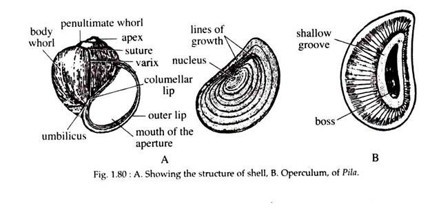
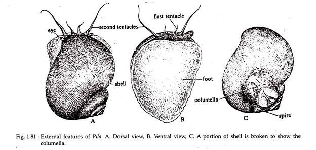
The extreme top of the shell is called the apex (Fig. 1.80A), which is regarded as the oldest part of the body. Starting from the apex the other whorls — the penultimate whorl and body whorl are large so as to enclose the greater part of the body. The first whorl is smallest and the last one is largest (Fig. 1.81A).
The last whorl contains a large aperture, which can be closed by a lid called operculum (Fig. 1.80B), which is attached to the posterior side of the foot. The operculum is a flat calcareous plate, formed as a cuticular secretion of a group of cells from the foot. The operculum has a lunate oblong outline which corresponds to the aperture of the shell. It shows numerous concentric ring of growth around a well-marked nucleus.
The inner surface of the operculum shows a distinct elliptical area called boss (Fig. 1.80B) for the insertion of opercular muscles. The margin of the aperture is smooth and is called peristome. A spiral column arising from the centre of the shell is present on the inner side called columella (Fig. 1.81C).
The type of coiling seen in Pila globosa is right handed and is called dextral, which is in contrast to the other rare type of left-handed coiling, called sinistral.
ADVERTISEMENTS:
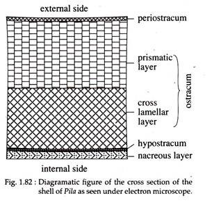
The microscopic structure of the shell of Pila shows three distinct layers. The outermost layer is chitinous and known as periostracum, while the two underlying layers, the ostracum and hypostracum, are composed of calcareous material. The periostracum is thin and consists of a large number of parallel bands in young stage. Each band is made up of rectangular blocks and are separated from one another by wavy lines.
In adults, the periostracum, however, appears as a homogenous membrane without showing the bands or blocks. The ostracum is composed of two distinct layers — Prismatic layer and cross lamellar layer (Fig. 1.82). These layers are calcareous in nature. Hypoostracum is a thin calcareous layer. The innermost layer is nacreous layer, which is very smooth and glossy.
The whole body is located within the whorls of the shell. The columellar muscle arising from the foot attaches the body to the columella. The columellar muscle plays a vital role. It prevents the animal from extending out of the shell beyond certain limit and also helps to withdraw it into the shell. The head along with the foot can be protruded to a limited extent through the aperture of the shell.
The body of Pila is divisible into the head, foot and visceral mass. Head is well marked and prolonged into a partly contractile snout. It carries two pairs of tentacles. The longer pair is filament-like, hollow and contractile. At the base of each tentacle projects a small, stumpy eyestalk or ommatophore bearing a prominent bead-like eye at its tip.
The shorter pair of tentacles is called labial palps or first tentacle and is regarded as the anterio-lateral prolongation of the snout. Two fleshy projections, called nuchal lobes or pseudoepipodia, are seen on the two sides of the head.
The lobes projecting anteriorly over the foot, are the prolongations of the mantle and are innervated by nerves from the pleural ganglia. The left nuchal lobe is highly developed and forms the respiratory siphon.
The foot is large, strongly muscular and is more or less triangular when seen from the ventral side (Fig. 1.81B). The anterior part of the foot is round and the posterior part of the foot holds the operculum. The foot of Pila has a broad, flat, smooth ventral surface, the sole, adapted for creeping movement. It is highly muscular and contains pedal glands.
The visceral mass forms a sort of hump on the dorsal side and contains all the main organs of the body. It is spirally coiled like the shell and exhibits the phenomena of torsion. The skin covering the visceral mass forms the pallium or mantle.
ADVERTISEMENTS:
The mantle subserves three functions in the life of Pila:
(i) Protects the visceral mass and head,
(ii) Serves as an additional respiratory organ and
(iii) Secretes the shell with the help of the shell-secreting nacreous glands at the free margin of the mantle.
The mantle is free anteriorly and encloses a spacious cavity known as pallial or mantle cavity, which houses visceral organs of the animal. The mantle cavity is imperfectly divided into left and right chambers by a longitudinal ridge known as epitaenium.
The right chamber contains the ctenidium, rectum and the genital duct. The left chamber contains the pulmonary sac. There is a comb-like, sensory structure known as osphradium close to the left nuchal lobe. The mouth and anus are closely situated on the same side of the body. The anal and the genital apertures are located on the right mantle opening.
4. Integumentary System of Pila:
The skin greatly varies in thickness. It consists of an outer epidermis and an inner, more complex dermis. The epidermis covering the whole body consists of a single layer of epithelium, some of which are modified into unicellular glands, from which mucus, pigment and lime are secreted. The dermis or corium includes muscle fibres and connective tissue and is quite rich in pigment cells.
5. Body Cavity of Pila:
The general body cavity in the adult, is haemocoel. The true coelom is greatly reduced and is represented by the pericardial cavity and the cavities around the kidney.
6. Locomotion in Pila:
The foot of Pila helps in locomotion. The flat sole of the foot helps Pila to move very slowly by creeping on the substratum. During movement the foot is protruded through the opening of the shell and this extension is brought about by the sudden influx of blood into it. The glands present in the foot produce a slimy secretion that helps the animal to glide on dry surface.
The foot is provided with vertical, longitudinal and transverse muscles. During locomotion the wave-like contractions on its surface are produced by the contraction of the vertical muscles. The contraction of the transverse muscles drives the blood forward which causes the extension of the foot in front. During this process the longitudinal muscles contract to pull the posterior end of the foot forward.
7. Digestive System of Pila:
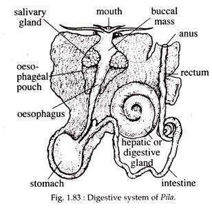
Pila is herbivorous and it lives primarily on aquatic vegetation. Its digestive system comprises of a tubular digestive canal and digestive glands (Fig. 1.83).
Digestive canal is made up of three distinct regions: (i) fore gut, (ii) mid gut and (iii) hind gut. The fore gut and hind gut develop from the embryonic ectodermal layer, while the mid gut is endodermal in origin.
(i) Fore Gut:
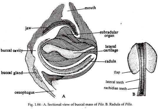
The fore gut includes the buccal mass and the esophagus. Mouth is a vertical slit which leads into the anterior end of the digestive tract which becomes greatly swelled to form an oval buccal cavity. The buccal cavity is enclosed by a strong thick- walled muscular structure called buccal mass. Many workers consider the buccal mass as the pharynx.
The entrance of the mouth is guarded by a pair of chitinous jaws projecting from the roof of the buccal cavity (Fig. 1.84A). At the floor of the buccal cavity, is present a chitinous ribbon-like structure called radula or lingual ribbon (Fig. 1.84B).
It is an elongated structure bearing transverse rows of serrations. Each transverse row contains about seven teeth — two marginals and a lateral on either sides of a median rachidian tooth, forming the formula 2, 1, 1, 1, 2 = 7. The radula is movably placed by muscles upon a large outgrowth of the floor of the buccal cavity called tongue mass or odontophore, which is made up of muscle with cartilaginous support.
The odontophore has an anteriorly placed subradular organ, which is more or less a rounded structure. It is divided into two by a median furrow. A small, pouch-like sublingual cavity is present beneath the subradular organ. The radula at the posterior end enters into a radular sac which supplies new teeth to the radula.
The radula is pushed forward by muscles from behind and it works as a file for rasping food materials. Dorso-laterally in the anterior region of the roof of the buccal mass lies a pair of jaws. The jaws are flexible.
Its anterior cutting edge is truncated and serrated, bearing numerous small and two or three large teeth-like processes. The jaws help to cut the aquatic vegetation upon which Pila feeds. The buccal cavity receives the secretion of two salivary glands, situated on its posterior side.
The buccal cavity leads into a long, narrow oesophagus. The oesophagus, just after its origin from the buccal mass, gives out on each side, small outpushings called oesophageal pouch. The oesophagus leads into the stomach.
(ii) Mid Gut:
The mid gut consists of the stomach and the intestine. It is red in colour and is situated on the lower part of the visceral mass just below the pericardium. It is a large sac, bent on itself to form a ‘U’-tube, one limb of which receives the oesophagus and the other leads into the intestine.
The portion at which the oesophagus ends is called the cardiac chamber, while the other end is called the pyloric chamber. The cardiac chamber constitutes the main part of the stomach. The wall of the cardiac chamber is corrugated while that in the pyloric part exhibits transverse folds.
The pyloric stomach is followed by a long narrow intestine that forms 2.5-3 coils. The posterior part of the intestine is nearly straight and turns to the anterior direction when it ends into the rectum.
(iii) Hind Gut:
It includes the rectum which is a thick-walled tube. It lies on the floor of the right side of the mantle cavity and finally opens to the exterior through a small anus. Anus is situated near the mouth within the right mantle opening.
Digestive glands include the salivary gland and the hepatic gland/digestive gland liver. There are two salivary glands situated on each side of the oesophagus. The two ducts of the salivary glands run anteriorly to open into the buccal cavity.
Their secretion consists of mucus and a starch digesting enzyme. The hepatic gland is black in colour and constitutes the main bulk of the visceral hump. It gives out two ducts which unite to form a common duct that opens into the stomach.
8. Respiratory System of Pila:
Pila is amphibious in nature and exhibits double mode of respiration. It is capable of absorbing oxygen dissolved in water by ctenidia or gills and is also capable of utilizing atmospheric oxygen by the pulmonary sac.
9. Circulatory System of Pila:
The circulatory system of Pila is well developed and has attained great complexity due to its double mode of respiration, Involving a gill as well as a lung. The circulatory system consists of the heart, the pericardium, the arteries, the veins and the sinuses.
The pericardium is a thin-walled roughly ovoidal chamber lying dorsally on the left side of the body and extending anteriorly up to the stomach and the digestive gland. The pericardial chamber encloses the heart and the aortic ampulla.
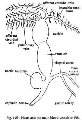
The heart is situated in the left-hand side of the visceral whorl very near to the posterior end of the ctenidium. As the ctenidium lies in front of the heart, the animals are included under Prosobranchia. The heart consists of two chambers, an auricle and a ventricle (Fig. 1.85).
The auricle is a thin- walled, highly contractile and roughly triangular sac situated in the dorsal part of the pericardium. The dorsal part of the auricle receives blood from three main veins — ctenidial vein, branchiorenal vein and pulmonary vein. Ventrally the auricle communicates with the ventricle through the auriculo- ventricular aperture.
The auriculo-ventricular aperture is guarded by semilunar valves which prevent regurgitation of blood from the ventricle to the auricle. The ventricle is an ovoidal sac lying below the auricle. Its wall is thick, spongy and muscular. The auricle receives oxygenated blood from the ctenidium and pulmonary sac through efferent ctenidial and pulmonary vein, respectively.
The lower end of the ventricle gives rise to a large artery, the aortic trunk. The root of the aorta is provided with two semilunar valves which do not allow the backflow of blood into the ventricle. The aorta immediately bifurcates into two arteries, the anterior one is called cephalic aorta, supplying blood to the head region and the posterior or visceral aorta which supplies blood to the posterior part of the body.
The cephalic aorta, just after its origin, gives a dilated sac-like outgrowth known as aortic ampulla. Both the aortae supply arteries to different parts of the body.
The cephalic aorta, along its outer side, gives off three arteries:
(i) an artery to the skin,
(ii) an artery to the oesophagus and
(iii) an artery to the left part of the mantle, the osphradium and left siphon.
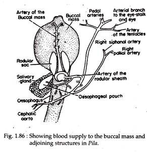
The cephalic artery on its inner side gives off a pericardial artery that supplies to the pericardium and then enters into the posterior renal chamber. The main trunk of the cephalic artery enters into the perivisceral sinus (space surrounding the buccal mass and oesophagus) and then crosses beneath the oesophagus.
It then gives off many arteries to the buccal mass, oesophageal wall, right side of the mantle, right siphon, copulatory organ, eyes, tentacles etc. (Fig. 1.86).
The visceral aorta, immediately after its origin, gives off an artery to supply the pericardium, digestive gland and skin. A little further, the visceral aorta gives rise to a stout gastric artery to the stomach. The main visceral artery runs along the left margin of the posterior renal chamber and sends branches to the intestine and the posterior renal chamber.
It then sends an artery to the digestive gland, the gonad and terminates in the wall of the rectum.
The blood, after being distributed to the various parts of the body by the arteries and their tributaries, passes into small spaces (lacunae). These lacunae unite to form large sinuses.
There are four main sinuses:
(i) peri-visceral sinus,
(ii) peri-intestinal sinus,
(iii) branchio-renal sinus and
(iv) pulmonary sinus.
The perivisceral sinus sends blood to the ctenidium and pulmonary sac. The peri-intestinal sinus passes blood to the kidney for eliminating metabolic waste.
The veins carry blood to the auricle from different parts of the body either directly or through the gill, mantle and kidneys.
The main veins are:
(i) afferent ctenidial vein,
(ii) efferent ctenidial vein,
(iii) afferent renal vein,
(iv) efferent renal vein and
(v) pulmonary vein.
Blood:
The blood of Pila contains some colourless stellate amoeboid cells and a blue, copper containing respiratory pigment, called haemocyanin. The amoeboid cells are phagocytic in nature.
Circulation of Blood:
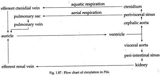
The ventricle of the heart pumps blood through the branches of cephalic aorta and visceral aorta (Fig. 1.87). The cephalic and visceral aortae supply blood to the different parts of the body. The cephalic aorta supplies blood to the head, mantle, buccal mass, oesophagus, copulatory organ, columellar muscle and associated structures.
The visceral aorta supplies blood to the visceral mass. Although there are four main sinuses, the blood is collected into the perivisceral and peri-intestinal sinuses. From these sinuses blood is conveyed either into the pulmonary sac, ctenidium or into the kidney.
During aerial respiration, blood flows into the pulmonary sac, while in aquatic respiration most of the blood from the perivisceral sinus goes to the ctenidum (Fig. 1.87). After purification, the blood comes to the auricle by the pulmonary vein or by the efferent ctenidial vein. The blood from the peri- intestinal sinus passes either into the anterior or posterior chamber of the kidney.
On its way through the anterior renal chamber, the blood gets rid of nitrogenous waste and flows either into the ctenidium or into the posterior renal chamber. The posterior renal chamber gets blood either from the peri- intestinal sinus or from the anterior renal chamber. The blood gets rid of its excretory product but without being aerated. Thus mixed blood goes to the auricle for distribution via the ventricle.
10. Excretory System of Pila:
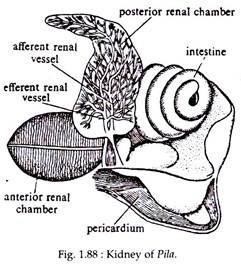
The excretory organ in Pila is the kidney. It consists of two renal chambers — one anterior and the other posterior (Fig. 1.88). It is coelomic in origin, being a true coelomoduct, opening at one end, into the coelom (pericardial cavity) and at the other, to the exterior (mantle cavity).
The anterior renal chamber is more or less oval in shape, lying in front of the pericardium in the hind part of the mantle cavity. It communicates with the posterior renal chamber by one end while the other end opens into the mantle cavity through a slit-like aperture near the epitaenia.
It is reddish in colour and its internal cavity presents numerous lamellated processes which reduce the internal cavity on the floor, the lamellae are arranged on either side of the afferent renal sinus, while on the roof they are similarly arranged on either side of the efferent renal sinus.
The posterior renal chamber is broad and the colour varies from brownish to grey. It is situated behind the anterior renal chamber, in between the rectum on the left and the pericardium and the hepatic gland on the right. Its large, internal cavity encloses a part of the genital duct and a few coils of the intestine.
This posterior renal chamber is separated from the pericardium by a vertical partition (renopericardial septum) and opens into it through a slit-like renopericardial aperture. The roof has profuse branching of the afferent and efferent renal vessels.
The two renal chambers are provided with network of blood vessels and take up nitrogenous waste from the blood. The excretory fluid from the posterior chamber is transferred to the anterior chamber, from where the waste is discharged into the mantle cavity through the renal duct.
From the mantle cavity the waste products are finally eliminated out of the body through the right siphon along with the out flowing water.
The excretory fluid contains mostly ammonia, urea and uric acid. Pila being amphibious shows an adaptation for water conservation during terrestrial phase by converting ammonia into the insoluble uric acid. It, for example, has 1.68 mg of uric acid per gram of kidney when freshly collected, but comes to have up to 102 mg uric acid per gram of kidney when it is aestivating under dry conditions.
11. Nervous System of Pila:
The nervous system of Pila comprises of ganglia, commissures, connectives and nerves to different part of the body. Pila also has sense organs such as osphradium, tentacles, statocyst and eyes. Reproductive
System:
Pila is dioecious but sexual dimorphism is poorly marked. The male has a much more developed copulatory organ than those of female.
Male Reproductive System:
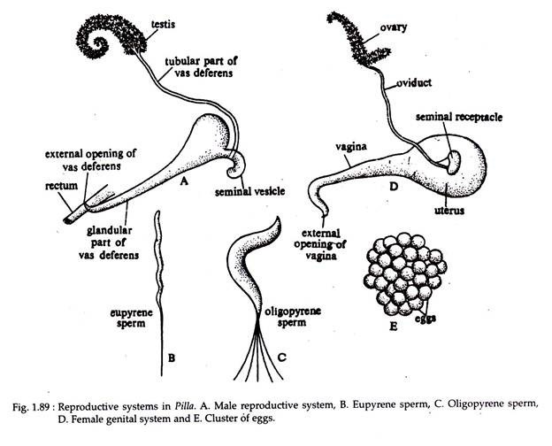
The male reproductive system comprises of testis, vasa efferentia, vas deferens, vesicula seminalis, penis and hypobranchial gland (Fig. 1.89A).
The testis lies in close contact with the hepatic gland and occupies the upper two or three whorls of the shell. The testis is a single, flat, plate-like structure and is more or less triangular in outline. It is cream in colour. Many fine ducts the vasa efferentia, originate from the testis, unite together to open into the vas deferens.
The vas deferens or male gonoduct is differentiated into:
(i) an upper tubular part,
(ii) a terminal glandular part and
(iii) a curved blind tube called vesicula seminalis.
The upper tubular part is narrow and thin walled. The terminal glandular part is thick-walled which runs forward on the left side of the rectum. It finally opens by the male genital aperture.
The vesicula seminalis is a somewhat curved, swollen structure with a blind, rounded posterior prolongation and situated between the junction of the two parts of the vas deferens. It serves as a storehouse of the sperms. The penis is present in the form of a whip-like flagellum which is partially ensheathed by the penis sheath and situated on the right side of the body near the mantle opening.
The penis sheath is a simple outgrowth from the inner surface of the mantle. The penis is capable of great extension. At the base of the penis sheath is an oval glandular thickening, the hypobranchial gland, consisting of tall cells containing small basal nuclei.
Two types of spermatozoa are encountered in Pila:
(i) Eupyrene type which is pear shaped and
(ii) Oligopyrene type which is worm like (Fig. 1.89B & C).
The eupyrene type is functional and is capable of fertilizing the egg.
Female reproductive system. The female reproductive system consists of ovary, oviduct, receptaculum seminalis, uterus, vagina and albumen gland (Fig. 1.89D).
The ovary in female Pila occupies the same position as the testis of the male, but is less extensive and lies towards the inner side of the hepatic gland. It is a much branched, orange coloured structure. Female gonoduct or the oviduct which is differentiated into an upper tubular part, comes down along the edge of the hepatic gland. The lower glandular part remains on the floor of the mantle cavity parallel to the rectum.
The glandular part is distinguishable into a yellow coloured albumen gland which secretes albumen and the posterior part is distinguished as the uterus. The uterus is large, yellow and pear shaped, lying in the body whorl below the intestine and to the right of the renal chamber.
At the junction of the tubular and glandular portion of the oviduct there is the receptaculum seminalis or the spermatheca where spermatozoa remain stored. The terminal part of the uterus is differentiated as vagina.
The vagina is a white or cream-coloured, band-like tube, running forward just beneath the skin. It opens by a narrow, slit-like aperture, a little behind the anus. The male intromittent organ, the penis is present in females which is rudimentary and functionless.
Few hundred eggs are laid at a time and they adhere together to form a mass (Fig. 1.89E). The eggs are fertilized by spermatozoa coming from the spermatheca. Fertilization is internal and oviposition begins a day or two after copulation. The zygote receives albumen and a coating of shell in the uterus. Existence of a rudimentary penis in female is suggestive of the fact that this group had hermaphroditic ancestor.
12. Development of Pila:
Development occurs outside the body of the female. The parents do not incubate or look after the eggs. Development of Pila is direct. The embryo floats in a central core of liquid albumen which is surrounded by a thick layer of whitish solid albumen.
During development the visceral mass and the shell of the embryo becomes spirally coiled and the characteristic phenomena of torsion takes place. This results in the asymmetry of the body. The young snails hatching from the eggs are similar in form to the adult.
