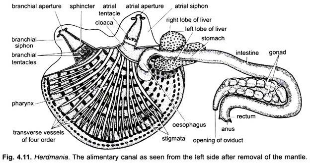
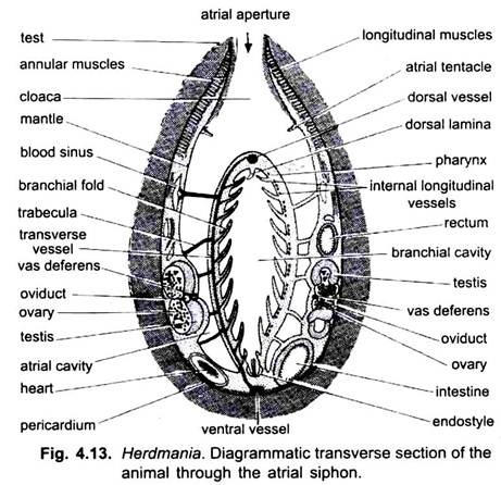
In Herdmania, digestive system includes the alimentary canal and digestive glands. Lying in the atrial cavity inside the mantle is a large bag-like pharynx or branchial sac. The wall of the pharyx is fused to the mantle along the anterior and ventral sides, on account of this attachment the atrial cavity is divided into right and left halves which are, however, continuous dorsally.
Alimentary Canal:
The alimentary canal is coiled, beginning from the mouth and ending in anus. It has the following parts:
1. Mouth:
The mouth or branchial aperture lies at the top of branchial siphon, which is designated as the anterior end of the body. It is guarded by four lips (lobes) of the test. The mouth leads into a branchial siphon.
ADVERTISEMENTS:
2. Branchial Siphon:
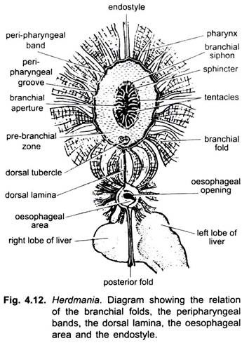
It is a narrow and tubular cavity lined by ectoderm and is called the stomodaeum or buccal cavity. At the base of the stomodaeum is a ring of branching tentacles which act as a sieve allowing only minute food particles to go in. These are richly innervated and their number is about 64.
Below the tentacles is a smooth prebranchial (prepharyngeal) zone having a swollen dorsal tubercle made of two spiral coils. Then there are two thin ciliated ridges, the peripharyngeal bands lying parallel and encircling the upper end of the pharynx. The posterior one of the two peripharyngeal bands is joined mid-dorsally to a dorsal lamina, and mid-ventrally with an endostyle.
3. Pharynx:
ADVERTISEMENTS:
It occupies the major part of the body cavity and is differentiated into two- prebranchial zone and branchial sac.
(i) Prebranchial Zone:
It is the smaller anterior region having smooth walls without folds, cilia and stigmata or gill-slits.
ADVERTISEMENTS:
(ii) Branchial Sac or Pharynx:
It is the posterior larger part of the pharynx. The pharynx almost fills the atrial cavity. The wall of the pharynx is perforated by several rows of stigmata arranged in transverse rows, through these the cavity of the pharynx communicates with the atrial cavity. The edges of stigmata are beset with cilia which drive currents of water from the pharynx into the atrial cavity.
The stigmata are not gill-clefts, but they are formed by sub-divisions of a few original gill-clefts. Between the stigmata the wall of the pharynx has transverse and longitudinal bars containing blood vessels, thus, the pharynx appears like a basket. The lining of the pharynx is raised into ten longitudinal folds on each side.
(a) Dorsal Lamina:
Dorsal lamina is a thin flap lying inside the mid-dorsal roof of the pharynx. It bears a number of conical, ciliated projections called languets. The dorsal lamina runs from the posterior peripharyngal band to the opening of the oesophagus. The dorsal lamina and its languets can bend to one side to form a sort of tube through which food-laden mucus passes.
(b) Endostyle:
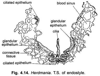
Lying in the mid- ventral floor of the pharynx is an endostyle. It is a longitudinal groove with four longitudinal rows of gland cells with ciliated cells between them. The middle row of cells has long cilia. The gland cells of endostyle secrete mucus which is driven laterally by ciliated cells. Endostyle is homologus with thyroid gland of vertebrates.
The posterior region of the pharynx is known as oesophageal area, around which all the folds of the pharynx converge. It has two semi-circular lips enclosing an aperture which leads into an oesophagus.
4. Oesophagus:
ADVERTISEMENTS:
It is a short bent tube having four ciliated groves in its lining which direct the food into the stomach.
5. Stomach:
The stomach is wider than the oesophagus but its walls are thin and have a tubular, branching pyloric gland. The tubules of the pyloric gland open by a single aperture in the middle posterior fold of the intestine. Stomach opens into a thin-walled intestine.
6. Intestine:
It has two parallel limbs forming a U. The intestine joins a short ciliated rectum opening by an anus into the dorsal part of the atrial cavity known as cloaca. The cloaca leads dorsally into the atrial siphon, opening outside through atriopore or atrial aperture. Like branchial siphon, the atrial siphon is also lined by ectoderm and, thus, represents the proctodaeum.
Digestive Glands:
1. Liver:
Lying against the stomach is a dark brown bilobed liver, the left lobe being larger. The liver is formed of a large number of fine tubules embedded in the matrix of connective tissue having blood sinuses. These tubules unite to form 11 or 12 hapatic duct which open separately into the stomach. The hepatic secretion has a powerful amylase, protease and weak lipase. Bile pigments are also present. In the liver and stomach are starch-like granules which are probably stored food.
2. Pyloric Gland:
It is located in the walls of the posterior part of stomach and intestine. Pyloric gland is branched and its ductules unite to form a common duct which opens in the intestine. Its secretion is pancreatic in nature. It also probably acts as an excretory organ.
Food, Feeding and Digestion:
(i) Food:
Microorganisms such as protozoans, pieces of decaying animals, zooplanktons and algae are its food.
(ii) Feeding:
Ciliary feeding takes place because of sedentary habit. Cilia bordering the stigmata keep up a constant current of water entering the mouth, and passing ciliated epithelium into the pharynx from where it goes out through the stigmata into the atrial cavity, and then to the exterior through the atrial aperture. The current of water carries in minute organic food particles which pass into the pharynx, where they settle down on the pharyngeal walls.
Gland cells of the endostyle secrete mucus (which is not carried forward by cilia as was originally believed). The mucus is lashed out transversely from the endostyle by its cilia, at right angles to the endostyle. Food particles lying on the lining of the pharynx are caught in the mucus and generally transported upwards along the pharyngeal wall to the dorsal lamina, in passing the food particles and mucus are rolled into a cylindrical mass. The dorsal lamina cilia convert the cylindrical mass into a string in the tube formed by curving upwards of languets. This food-laden mucus string is carried into the oesophagus and then into the stomach.
(iii) Digestion:
The liver secretes a yellowish-brown digestive fluid into the stomach; it has many enzymes, an amylase which splits carbohydrates into maltose, a protease which breaks down proteins, and a weak lipase which probably acts on fats. The liver also stores starch as reserve food material.
The pyloric gland is an accessory digestive organ; it is probably pancreatic in nature. Food is digested mainly in the stomach and absorption occurs in the intestine. The ciliated rectum expels the much-coiled excreta with such force that it is shot out about 10 cm through the atrial siphon.


