
The paired cranial and spinal nerves constitute the peripheral nervous system. The autonomic nervous system is a part of the peripheral nervous system that controls the involuntary functions of the body.
Cranial nerves:
The cranial or cerebral nerves are paired, originate in the nuclei in the brain, pass out of the cranium through passages in it and terminate in the effectors. The cranial nerves are ten pairs in fishes and amphibia and twelve pairs in amniota, i.e. reptiles, birds and mammals.
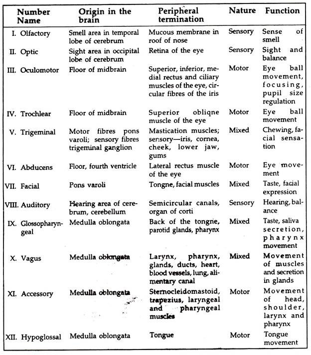
Some of the nerves are sensory, some are motor and others are mixed. The cranial nerves are referred to as nerves I to XII (Fig. 13.4). The number, name, origin in the brain, peripheral termination, nature and function of vertebrate cranial nerves are given in the table.

Spinal nerves:
Paired nerves arising from the spinal cord are known as spinal nerves. One pair of nerves from each somite emanate laterally from the spinal cord. Each nerve has two roots, the dorsal root bears a ganglion, the dorsal ganglion and a non-ganglionated ventral root (Fig. 13.5). Each root originates from one horn of the grey matter and the two join to form the trunk of the nerve.
The nerve comes out of the spinal canal, the space between the spinal cord and the vertebrae, through the passage between the arches of the adjacent vertebrae. The nerve roots have a covering of Dura and arachnoid maters, which terminate before the formation of the trunk.

The dorsal root consists of sensory neurons, carries sensory impulses to the spinal cord from the skin, body somite’s and viscera. Impulses are interrupted by a Synapse in the ganglion. The ventral root consists of motor neurons, carries impulses from the spinal cord to somatic and visceral components. The dorsal root may possess motor neurons but the ventral root never possesses sensory neurons.
Immediately after leaving the vertebral column each .spinal nerve divides into a posterior ramus and an anterior ramus. The posterior (dorsal) ramus innervates the dorsal part of the axial musculature, skin of the back and lateral and ventral parts of the skin and musculature of the body wall. The anterior (ventral) ramus innervates internal organs, smooth muscles, blood vessels and glands.
The visceral motor component enters a sympathetic ganglion. The sympathetic ganglia of one side are connected to a longitudinal sympathetic trunk and a trunk lies on each side of the vertebral column. The visceromotor system terminates in viscera, blood vessels, etc. and termed autonomic system. The visceromotor and viscerosensory systems together form the sympathetic nervous system.
Reflex action:
ADVERTISEMENTS:
Not all activities of animals are the results of judgement and deliberations. Certain actions occur without even the animal being aware of it, e.g. sudden withdrawal of hand or feet from hot, cold, sharp and pointed objects; sudden closure of eyelids in strong light, etc. Such actions are termed reflex actions. These are protective or inherited reflexes (present at birth).
Reflex actions which were not initially present but developed due to constant practice for a long time or learning by habit are known as conditioned reflexes. Walking, cycling, secretion of saliva in dogs with the ringing of a bell at feeding time, etc. are conditioned reflexes.
Reflex arc:
The neural pathway along which the impulses travel to execute the reflex action is called reflex arc. Each spinal nerve has two roots. Sensory neurons are present in the dorsal root and only motor neurons are present in the ventral root. In a simple reflex arc (Fig. 13.5) the impulse received from the receptor passes to the spinal cord through afferent neurons via the dorsal root ganglion and dorsal root.
The impulse from the spinal cord passes to the motor neurons in the ventral root via the synapse between the two and transmitted to the effector. The knee jerk is a typical simple reflex. One or more interneurons between the sensory and motor neurons are involved in the majority of the reflexes. Other reflexes are righting reflex and anti- gravity reflex.
Autonomic nervous system:
The part of the nervous system controlling the functions of certain organs in an animal body, automatically or involuntarily, is known as autonomic nervous system.
A number of organs, viz. eye, nose, heart, lung, gut, ciliary and iris muscles of the eye, muscles related to hairs, glands, gall bladder, urinary bladder and many other organs are supplied with a special set of peripheral nerves made of self- operating units, and their functions cannot be controlled voluntarily.
The autonomic system differs from the rest of the nervous system in the following features:
ADVERTISEMENTS:
1. It has a double set of nerves, both sensory and motor.
2. The axon endings of each set secrete a different transmitter substance to the effectors.
3. Cerebrum has no voluntary control over.
The autonomic nervous system has two components, the sympathetic and parasympathetic. Sympathetic and parasympathetic systems usually have antagonistic effects on the organs involved:
1. Sympathetic nervous system:
A chain of sympathetic ganglia extending from the upper-cervical level to the sacrum, lie on each side of the vertebral column (Fig. 13.6). The afferent (sensory) nerves pass to the spinal cord through the dorsal root ganglion, and dorsal root.
The efferent (motor) neurons are long and send small preganglionic fibres not to the grey matter of the spinal chord but to the autonomic ganglion via a small branch of the segmental nerve known as ramus communicants.
In the ganglion the neurons link up with motor neurons which send postganglionic neurons to the effector. Sympathetic nervous system action resembles that of adrenaline and ephedrin and inhibited by ergo toxin.

2. Parasympathetic nervous system:
In parasympathetic system the preganglionic neurons are long and join the parasympathetic ganglion located on the organ concerned. The postganglionic neurons are short. The action of parasympathetic nervous system resembles the action of acetylcholine and inhibited by atrophine.
The sympathetic nervous system increases heart rate, blood pressure, dilates bronchioles reduces gastrointestinal motility and secretions, constricts gastrointestinal sphincters, dilates pupil, relaxes gall bladder and urinary bladder. The parasympathetic actions are opposite to those of the sympathetic system.