In this article we will discuss about the Respiration in Cockroaches:- 1. Respiratory Structure in Cockroach 2. Mechanism of Respiration in Cockroach.
Respiratory Structure in Cockroach:
Spiracles:
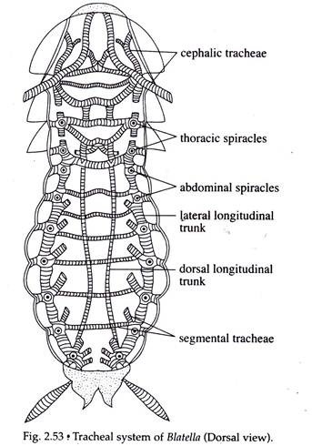
Ten pairs of spiracles or stigmata are present on the lateral side of the body. The largest first pair is present on the mesothorax. The second pair is on the metathorax and the rest eight pairs are on the first eight abdominal segments. Each spiracle is oval in shape and bounded by an annular sclerite having a filtering apparatus formed by the bristles to eliminate dust particles from the inflowing air.
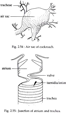
The spiracles in mesothorax have two lips — a rigid anterior lip and a movable posterior lip. In metathorax, the lips of the spiracles are united ventrally. In abdomen, the spiracles have no lips. The thoracic spiracles open directly within the segmental trachea, but the abdominal spiracles open first within the dilated part of the trachea — the atrium, from which originate the segmental tracheae.
ADVERTISEMENTS:
Tracheae:
In Blatella, the haemocoel contains a network of elastic, closed air tubes or tracheae. Three longitudinal tracheal trunks are present on each side of the abdominal cavity.
The dorsal and ventral trunks are present near the middle line, while the lateral trunk is present on the lateral side of the abdominal cavity (Fig. 2.53) Each lateral trunk is divided into two parts — the anterior part is present between mesothoracic, metathoracic and the first abdominal spiracle, while the posterior part extends from the second abdominal spiracle to the eighth abdominal spiracle.
Each dorsal and ventral tracheal trunk originates from a trachea arising from the first abdominal spiracle. They extend up to the segmental branch arising from the eighth abdominal spiracle. Six tracheae originate from each mesothoracic spiracle which supply to the head, prothorax and mesothorax. From the remaining spiracles three segmental tracheae are given out on each side (Fig. 2.53).
ADVERTISEMENTS:
The longitudinal trunks and the segmental tracheae are swollen at several places and are known as air sacs (Fig. 2.54). Large tracheae are internally supported by spiral ring of chitin, called taenidia or intima (Fig. 2.55), which prevent the tracheal tubes from collapsing. In addition, chitinous fibrils of 10 to 30 nm thickness and an epicuticle of lipoprotein, line the lumen of the tracheae.
Tracheoles:
ADVERTISEMENTS:
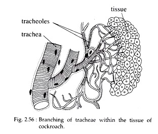
The branched tracheae anastomose and penetrate to all parts of the body of cockroach. The ultimate finer branches of tracheae are called tracheoles which come in direct contact with the individual cell (Fig. 2.56). They have a diameter of only 1 micron. Their walls are very thin and devoid of taenidia and other chitinous structures.
Instead, they are lined by a protein called trachein. The opening of each tracheole within the tissue is immersed within the body fluid which conveys respiratory gases to the cells. Thus, the elaborate tracheal system carries oxygen directly to all body cells.
Mechanism of Respiration in Cockroach:
Alternate contraction and relaxation of the abdominal targo-sternal muscles bring about changes in the diameter of the tracheae, so that air is forced in and out of the tracheal tubes through the spiracles. According to one view, during intake of air, the muscles relax to open the anterior four pairs of spiracles, through which air rushes in and reaches up to the intracellular spaces through the tracheoles.
During expiration, the abdominal muscles contract to drive the air out of tracheal space through the last six pairs of spiracles. According to another view, air flows in and out through all spiracles and probably there is no direct circulation of air along the longitudinal tracheal trunks.
The oxygen from the air dissolves in the fluid of the tracheoles and diffuses inwards to the tissues, in exchange for the carbon dioxide formed in them. During increased metabolism (at the time of flight), the amount of fluid in the tracheole is reduced by osmotic movement of water to the surrounding tissues.
This exposes more of the surface walls of the tracheoles to oxygen, causing increased oxygen supply to the tissues.
The working of spiracles is under the control of central nervous system. Cockroaches can close all the spiracles and may suspend its respiration for a considerable period of time. The opening aid closure of the spiracles depends upon the carbon dioxide concentration. The width of the spiracular opening increases with the rise of temperature from 20° to 32°C.
In cockroaches, blood takes no part in respiration, and the oxygen bearing fluid of the tracheoles serves in internal respiration, like that of lymph in vertebrates. In these insects more than 10% gaseous exchange can occur through body surface.


