
In this article we will discuss about Aurelia (Jelly Fish):- 1. Introduction of Aurelia 2. Structure of Aurelia 3. Histology 4. Musculature and Locomotion 5. Digestive Cavity and Canal System 6. Nutrition 7. Sense Organs 8. Life-history.
Contents:
- Introduction of Aurelia
- Structure of Aurelia
- Histology of Aurelia
- Musculature and Locomotion in Aurelia
- Digestive Cavity and Canal System in Aurelia
- Nutrition in Aurelia
- Sense Organs of Aurelia
- Life-history of Aurelia
1. Introduction of Aurelia:
Aurelia is most common of the larger jelly fishes. It is often found on the seashore, as a gelatinous saucer-shaped umbrella, several inches in diameter. In Amelia, medusa is the dominant and conspicuous zooid, while polypoid form is restricted to a short larval stage. The species discussed in this text is Amelia aurita which is popularly known as moon jelly.
Habit and Habitat:
ADVERTISEMENTS:
A. aurita is a cosmopolitan jelly fish. It occurs in the warm and temperate seas and lives in coastal waters singly or in large shoals. It is generally seen floating with the water current or swimming feebly by the contractile movement of its body.
It is carnivorous and feeds on small organisms. They display distinct photo-taxis. During cloudy weather and at twilight, they come to the surface, while during bright sunlight and at night, they move downwards.
2. Structure of Aurelia:
ADVERTISEMENTS:
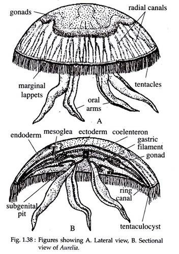
Adult Aurelia is like a large version of the medusa of Obelia (Fig. 1.38). However, the concave and convex sides of the umbrella are more pronounced. The jelly like transparent umbrella shaped body has four, red or purple horse-shoe shaped gonads on its upper surface and form long and narrow oral lobes hanging downwards from lower surface. The circular body of A. aurita measures about 90 mm in diameter.
The outline of the umbrella is somewhat circular, but is broken by eight equidistant notches, each bearing a pair of delicate sense organs, the marginal lappets or rhopalial lappets. At the centre of two rhopalial lappets is present the rhopalium covered by a hood.
Between the pairs of lappets the edge of the umbrella is provided with numerous closely set marginal tentacles. A narrow region of the umbrella, close to the edge, is very thin and flexible. This is the velarium bearing the marginal notches and the fringe of marginal tentacles. The velarium, quite unlike the true velum of the medusa of Obelia, contains gastro-dermal canals.
At the centre of the sub-umbrella surface is the mouth, which is distinctly four- cornered and is borne at the end of an extremely short and inconspicuous manubrium. The corners of the mouth are drawn out into four lobes called oral arms. Each oral arm consists of a folded membrane, tapering to a point at its distal end and containing minute lobules along its edges.
ADVERTISEMENTS:
The oral arms are abundantly provided with stinging capsules or nematocysts. The corners of the mouth and the arms lie in the four per-radii, which are the major body axis. Half way between the per-radii are the inter-radii and in-between the per-radii and inter-radii are the eight adradii (Fig. 1.39).
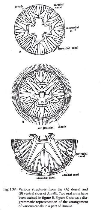
At a short distance from the mouth, at each inter-radial area, there is a somewhat circular aperture leading into a shallow pouch, the sub-genital pit (Fig. 1.39B). It lies immediately beneath one of the gonads. The sub-genital pits are not connected with the reproductive system. Gonads are four in number, situated at the floor of the gastric pouches.
The gonads are coloured, horse-shoe shaped with a frill like structure and its concavity facing towards the mouth. In contrast to the usual ectodermal gonad in hydrozoans, the gonads in scyphozoans are endodermal. The gametes, when mature, break into the gastro-vascular cavity and pass to the outside through the mouth.
3. Histology of Aurelia:
Histologically, the scyphozoan medusa has the same organisation as that of Obelia. The main difference lies in the structure of the mesoglea which contains fibres and loose amoeboid cells. Nematocysts are of three types — atrichous isorhiza, holotrichous isorhiza and heterotrichous micro-basic euryteles (Fig. 1.40). They occur on the oral arms, ex- and sub-umbrella surfaces, marginal tentacles and gastric filaments.

4. Musculature and Locomotion in Aurelia:
The muscular system in Aurelia is well developed. The broad circular muscle band on the sub-umbrella surface is very strongly built and is known as coronal muscle. Longitudinal muscles are present in the tentacles, manubrium and oral arms.
ADVERTISEMENTS:
The coronal muscle helps Aurelia in its swimming movements, during which the ex-umbrella surface is kept upwards. Rhythmic contractions of muscles force water out from the sub-umbrellar cavity as a result the body is propelled in the opposite direction.
5. Digestive Cavity and Canal System in Aurelia:
Mouth leads into a short tube or gullet which opens into a centrally located stomach. Stomach is produced into four inter-radial gastric pouches extending up to the half-way between the centre and the edge of the umbrella. Numerous delicate gastric tentacles or phacellae are present on the floor of the gastric pouches.
These are composed of endoderm with a core of mesoglea and abundantly supplied with nematocysts. The function of these structures is to kill or paralyze the prey if they are taken alive into the stomach. From the periphery of the stomach as well as from the gastric pouches, sixteen radial canals originate, of which four are per-radials, four inter-radials and eight adradials (Fig. 1.39A).
Both per radials and inter-radials are branched, while the adradials are un-branched. All the radial canals ultimately open into a ring or circular canal situated at the margin of the body.
6. Nutrition in Aurelia:
The nematocysts in jelly fishes are relatively powerful and they are capable of feeding on fishes and other rather larger preys. Aurelia is carnivorous and flagellary-mucus feeder. Small food particles and plankton are caught in slime secreted on the ex-umbrella.
These, slime entangled prey are then licked off the lappets by the oral arms. The particles are then carried into the stomach by the current produced by flagella in grooves on the oral arms. Aurelia feeding on larger prey use the gastric tentacles to pull a victim into the gastric cavity and if necessary paralyze it with nematocysts.
Moreover, the filaments contain many gland cells that secrete enzymes for preliminary digestion. Partly digested food particles, circulate through the canal system and are ingested by endodermal cells for intracellular digestion in food vacuoles. The outgoing current of water carries away the undigested material out of the body.
7. Sense Organs of Aurelia:
The sense organs of Aurelia are the tentaculocysts or rhopalia. The rhopalia are located around the bell margin between lappets. Each rhopalium is flanked by a pair of rhopalial lappets. They are housed in a little hollow club-shaped structure and is covered by a hood (Fig. 1.41).
Each rhopalium contains two sensory pits lined with sensory epithelium, a statocyst and an ocellus. The endodermal lithocytes or statocysts contain heavy calcareous particles that weight the rhopalial tip. The function of the rhopalia is the initiation and control of the rhythmic swimming movements of the animal.

In connection with each rhopalia are two cavities, one on the ex-umbrella and the other immediately internal to the sense-club. These cavities are lined with sensory epithelium and are called olfactory pits.
8. Life-history of Aurelia:
Aurelia is dioecious and the gonads are endodermal. The unisexual medusae do not exhibit any external difference in their appearance i.e. no sexual dimorphism. The male medusa produces many sperms which pass out into the surrounding water through the mouth.
The eggs are lodged in pits on the oral arms and this temporary brood chamber is the site of fertilization and early development to the formation of planula larva. In the brood chamber, zygote through repeated divisions, attains the morula stage. Morula by accumulation of fluid in its interior, becomes a blastula, which is a closed sac with walls formed of a single layer of cells (Fig. 1.42).
An invagination from one end obliterates the cavity of the blastula and a new cavity is formed within the layer of cells. The new cavity opens to the exterior through a small opening, the blastopore. This stage is called gastrula which ultimately transforms into a hollow two-layered planula larva. In Aurelia, the transformation of the zygote into a planula larva takes two days.
The basic pattern of the planula larva is same as in Obelia, except that the blastopore is not completely closed and is represented by a mere slit. The planula larva swims about by means of the cilia of its ectodermal cells and after a brief period of free existence, settles down and fastens itself to the sea bottom by one pole. It then loses its cilia and metamorphoses into the hydra-tuba stage.

Metamorphosis of Planula:
The planula larva after fixation to a substratum forms a definite mouth at the free end and is now called the hydra-tuba stage. The mouth becomes square and is raised into a short manubrium. The aboral portion becomes narrowed into a distinct peduncle.
On two opposite sides of the mouth hollow processes grow out, forming the first two tentacles and soon two others appear at right angles to the first two. Thus, it is provided with four per- radial tentacles. Subsequently, four inter-radial and eight adradial tentacles appear.
Eventually the hydratuba possesses sixteen tentacles. At the same time the endoderm of the enteric cavity is produced into four longitudinal ridges, inter-radial in position and is called the gastric ridges or taenioles.
The ectoderm between the mouth and the circlet of tentacles become invaginated in each inter-radius to produce four narrow funnel-like depressions, the septal funnels or infundibula, sunk in the four gastric ridges.
The hydra-tuba remains unchanged for a long time, feeds and produces horizontal branches from which new hydra-tuba buds off. Finally it becomes segmented by transverse furrows and this stage is known as scyphistoma. The hydra-tuba and scyphistoma represent the hydroid phase of Aurelia.
Scyphistoma sometime multiplies by budding from stolons. After some time scyphistoma undergoes transverse fission at its oral end, a process called strobilation. It becomes divided by a series of constrictions which deepens until the polyp assumes the appearance of a pile of saucers, each with its edge produced into eight bifid lobes, four per-radials and four inter-radials.
Soon the process of segmentation gets completed and these saucer like bodies detach from the body column and turning upside down, begin to swim about as a small jelly fish called an Ephyra.
Ephyra Larva:
The ephyra larva is characterised by the possession of eight long bifid arms with deep notches. The ephyra has carried away with it a segment of the stomach with gastric ridges of the scyphistoma. This, during the process of constriction becomes closed at the ex-umbrellar side, while on the sub-umbrellar side it remains open and grows out to form a short manubrium.
On each gastric ridge appears a single gastric filament, which is soon followed by others. In the notches at the tip of the eight arms, tentaculocysts at this time, become recognisable. The spacious enteric cavity then gets continued into the eight arms in the form of wide radiating canals.
As the ephyra grows, the deeply notched adradial regions grow more rapidly than the rest. The notches become gradually filled up and the eight-rayed umbrella becomes nearly a circular disc. Four oral arms develop from the four corners of the mouth and the numerous marginal tentacles also develop. The ephyra thus gradually assumes the form of the adult Aurelia.
Metagenesis in Aurelia:
The sexual generation being represented by the adult Aurelia and the asexual generation by the hydra-tuba and scyphistoma. But instead of the medusa being developed as a bud on a branched colony as in Obelia, it is formed by the metamorphosis of an ephyra, developed as one of several transverse segments of a polyp (scyphistoma).
Therefore, the life history of Aurelia may be described as a metagenesis rather than an alternation of generations.