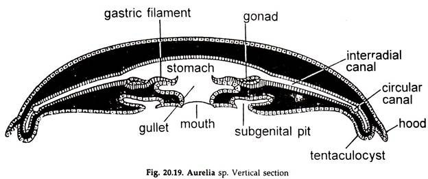
In this article we will discuss about the histology of aurelia, explained with the help of a suitable diagram.
a. The ectoderm covers the bell all around.
b. The gullet, and the sub-genital pits are also lined by the invaginated epidermis. The epidermis contains epithelial, sensory, nerve, gland cells and cnidocytes.
c. The cnidocytes occur on the oral arms, the ex- and subumbrellar surfaces, the marginal tentacles as well as on the gastric filaments.
ADVERTISEMENTS:
d. The gastro vascular cavity, its radiating canals and their extensions are lined by gastro dermis. The interspaces between the gastro vascular canals are filled with a thick solid layer of gastro dermal lamella. The cells are mainly flagellated columnar epithelial and gland cells.
e. Gonads are gastro dermal.
f. Nerve cells and muscle processes are wanting.
g. A thick gelatinous mesogloea, between the ecto- and endoderm, form the main mass of the umbrella. It contains branching fibres and wandering amoeboid cells derived from the ectoderm.
ADVERTISEMENTS:
h. The muscles are usually epidermal and striped, some are un-striped.
i. Simple un-striped longitudinal muscles form the musculature of oral arms and tentacles.
j. The muscles in the sub-umbrella are striated, and arranged in a strong, broad and circular band, called coronal muscle. The radial muscles extend along the main radii from the manubrium to the coronal muscle.
k. Locomotion is the result of rhythmic contraction of muscles.
ADVERTISEMENTS:
