In this article we will discuss about Obelia (Sea Fur):- 1. Introduction to Obelia 2. Structure of Obelia 3. Movement 4. Nutrition 5. Respiration, Circulation and Excretion.
Contents:
- Introduction to Obelia
- Structure of Obelia
- Movement in Obelia
- Nutrition in Obelia
- Respiration, Circulation and Excretion in Obelia
1. Introduction to Obelia:
Obelia is a typical example of a marine, colonial hydrozoan. Like most hydrozoans it has both polyp and medusa stages. Obelia occurs as a delicate whitish or light brown, almost fur-like growth on seaweeds or timber. It consists of branched filaments approximately the thickness of fine sewing cotton. A common example of it is Obelia geniculata.
ADVERTISEMENTS:
Habit and Habitat:
Obelia is typically marine, colonial and sedentary, having cosmopolitan distribution. It is found up to a depth of 80 metres. It exists in two principal form—the polyp and the medusa. The polyp represents the asexual phase which is a prominent branched hydroid colony found attached to rocks, stones, shells of animals, wooden pilings etc.
The medusa, on the other hand, represents the sexual phase. There is an alternation of these two phases in its life-cycle, which is a typical instance of metagenesis.
ADVERTISEMENTS:
ADVERTISEMENTS:
2. Structure of Obelia:
A. Hydroid or Polyp Phase:
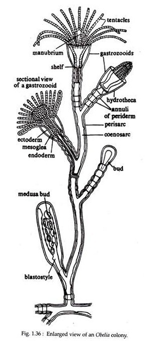
This stage of Obelia is a branched filament like structure and remains attached with the substratum. The colony is polymorphic, consisting of a number of individuals or zooids that are morphologically as well as functionally different from each other (Fig. 1.36).
i. Hydrorhiza and Hydrocaulus:
ADVERTISEMENTS:
The colony of Obelia consists of two portions — the horizontal portion called the hydrorhiza and the vertical portion, bearing the zooids, called hydrocaulus. Hydrorhiza is a branched structure and gives only mechanical anchorage to the whole colony. From the hydrorhiza arises the vertical hydrocaulus. Each hydrocaulus branches in an alternate manner.
The lateral branches may sometimes give off branches of a third order. Each branch bears zooid at its terminal end. Two types of zooids are seen — gastro-zooids having nutritive function and blastostyles for reproduction. In addition to the above two zooids, the lateral branches also bear short club-like structures which are either primordial or developing gastro-zooids or blastostyles.
Both hydrorhiza and hydrocaulus are hollow tubes called the coenosarc, covered externally with perisarc. Coenosarc is made up of two cellular layers, an outer ectoderm and an inner endoderm. In between these two cellular layers there is a thin non-cellular mesoglea or meso-lamella.
Coenosarc contains a tubular cavity, at the center known as coelenteron which is continuous throughout the colony. It is filled with a fluid. The tentacles of Obelia are solid and contain endoderm cells at its core.
The perisarc or periderm is a cuticle-like transparent non-cellular layer secreted by the ectoderm of coenosarc. Perisarc remains separated from coenosarc except at places where it shows attachment with it, thus showing some constrictions which are known as annuli of perisarc or periderm.
ii. Gastro-zooid or Tropho-zooid or Nutritive-zooid:
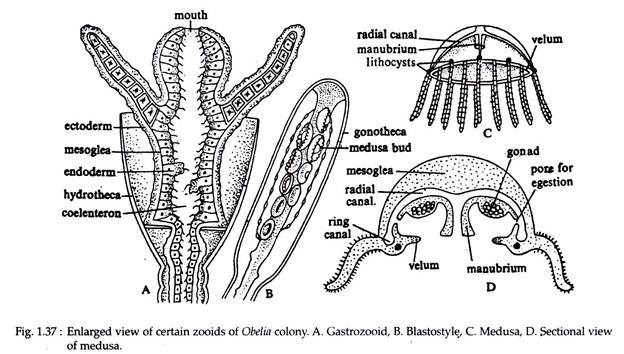
Majority of the zooids present in the hydroid stage of Obelia are the gastro-zooids. They are nutritive in function and are responsible for feeding the whole colony. Each gastro-zooid has a short tube-like body (Fig. 1.37A). At its distal end there is a conical projection called hypostome or manubrium.
Both body and manubrium are hollow, containing the spacious coelenteron. Mouth is situated at the terminal end of it. Surrounding the manubrium there is a circlet of about twenty four solid tentacles. This zooid is surrounded at the base by an investment called hydro-theca. Hydro-theca is transparent and is formed by the modification of the perisarc.
ADVERTISEMENTS:
It is shaped like a vase or wine glass. A short distance from its narrow or proximal end, it grows inwards into a circular shelf, upon which the base of the polyp rests. The circular shelf has a central aperture through which the tubular body of the zooid remains continuous with the rest of the colony. When irritated, the polyp undergoes contraction and suddenly withdraws, more or less, completely into the theca.
The mouth is capable of great dilation and contraction. Accordingly the manubrium appears either conical or trumpet shaped. Small organisms like aquatic crustaceans, nematodes and other worms are caught by the tentacles, armed with nematocysts. The food is then carried towards the mouth where they are swallowed.
iii. Blastostyle or Gonozooid or Reproductive-zooid:
These types of zooids are few in number in comparison to the number of gastro-zooids. Each blastostyle has a long cylindrical body without mouth and tentacles (Fig. 1.37B), but ends distally in a flattened disc. The coelenteron is greatly reduced. The hydortheca of a polyp is represented here by the gonotheca.
It is a cylindrical, transparent capsule enclosing the whole blastostyle. The lateral wall of the blastostyle gives off small lateral buds called the medusa-bud. They exhibit great structural variations depending upon the developmental sequences. When the medusa-buds become mature, the gonotheca ruptures at its distal end to allow the medusa-buds to escape.
iv. Histology of the Colony:
Histologically the structure of gastro-zooids, blastostyles and coenosarc are similar. They consists of two cellular layers — an outer ectoderm and an inner endoderm. In between the two layers there is a thin non- cellular mesoglea or meso-lamella.
Ectoderm is thin and chiefly composed of large, conical and columnar epitheliomuscular cells with their broad bases outwards. The compound name of this type is due to its dual functioning capacities, protection and the power of contraction.
The outer ends fuse and gives rise to a continuous cuticle along the entire length of the body. The narrower ends are directed to the mesogleal side and are produced into contractile processes. The contraction of these processes shortens the body column and tentacles.
This cell type has a prominent nucleus and several small vacuoles. Ectoderm also contains stinging cells or nematocysts, which are specially abundant on tentacles. Obelia possesses a single type of nematocyst, the basitricha. A nerve net composed of large and branched nerve cells is present on each side of the mesoglea.
Endoderm lines the gastro-vascular cavity throughout the colony. If consists chiefly of large, columnar nutritive-muscular cells. Nucleus is usually located towards the mesogleal side and contains one or two nucleoli. Cytoplasm is less vacuolated. Nutritive muscular cells are of two types — amoeboid and flagellated.
The broader end of amoeboid cells project in the coelenteron and produces pseudopodia to engulf food particles. The flagella of the flagellated cells, bring about a constant movement of food particles in the coelenteron. Among the nutritive-muscular cells are present small, narrow gland cells with granular protoplasm. They are responsible for secreting digestive enzymes.
B. Medusoid Phase:
Medusae are formed by the development of medusa-buds in the gonozooid and liberated from a ruptured gonotheca. When fully formed, it assumes the appearance of an umbrella with a convex surface by which the medusa was attached with the blastostyle (Fig. 1.37C).
This convex outer surface of the bell or umbrella is distinguished as the ex- umbrella and the concave inner surface as the sub-umbrella. From the surface of the subumbrellar surface emerges a hanging tube called manubrium bearing a square mouth at its terminal end (Fig. 1.37D).
As the medusa of Obelia swims, the umbrella becomes turned inside out, the sub-umbrella then forming the convex surface and the manubrium arising from its apex. This phenomenon is unknown in the medusa of other species.
At the edge of the umbrella arises a very short circular shelf called velum. There is a circlet of tentacles at the junction of exumbrellar side and velum. The number of tentacles is sixteen in a newly- formed stage.
The number may increase with age. At the bases of alternate tentacles there lies, a sense organ in the form of lithocyst or marginal sense organ. Each of these 8 lithocyst, is a minute globular sac containing a calcareous body in contact with hair-like processes of sensory cells. These sense organs regulate and co-ordinate the swimming movement of the organism.
The mouth leads into an enteric cavity or coelenteron, which occupies the whole interior of the manubrium. From the base of the coelenteron emerge four equidistant radial canals. They all open into a ring or circular canal, running at the margin of the body of the umbrella. By means of this system of canals, food taken into the mouth is digested in the manubrium and distributed to the entire body of medusa.
Histologically the structure of the medusa is same as that of the hydroid stage. The manubrium consists precisely the same layers — ectoderm, mesoglea and endoderm. Ectoderm is continued as the sub-umbrella and then round the margin of the umbrella on to the ex-umbrella.
The endoderm provides a lining to the whole canal system. In the umbrella, as in the manubrium, a layer of mesoglea is present between the two layers. Mesoglea is thick and often contains wandering cells or fibres. The velum, on the other hand, has a double layer of ectoderm, with a median layer of mesoglea. It has no extension of endoderm.
The tentacles, like those of the hydroid stage are formed of a core of endoderm covered by ectoderm. The cells of the ectoderm are abundantly supplied with stinging capsules. In the margins of both ex- umbrella and sub-umbrella, the ectoderm contains nerve cells. In ex-umbrella, the nerve cells crowd together to form an outer ring while in sub-umbrella it constitute the inner margin.
The whole body of medusa exhibits distinct radial symmetry. The medusae are unisexual (dioecious). Gonads are ectodermal in origin and remain in close association with the radial canals at the sub-umbrella surface. The gonads are round, four in number and externally both male and female gonads are similar.
3. Movement in Obelia:
Obelia colony being sessile, does not exhibit bodily movement from one place to another. They, however, undergo slight swaying movement under the influence of water current. The nutritive zooid can contract and expand their body and bend their tentacles considerably.
Medusa on the other hand can float passively in water and is drifted by the water currents with manubrium hanging downward and tentacles swaying freely. It can swim actively by muscular contraction. By rhythmic contraction and expansion, the water in sub-umbrella cavity is thrown backward and the medusa moves forward in a series of jerks.
4. Nutrition in Obelia:
Gastro-zooid or nutritive zooid is the feeding zooid of the Obelia colony. This zooid and medusa are carnivorous, feeding upon small aquatic crustaceans, nematodes and other worms. Their tentacles are armed with nematocysts, which paralyze and capture the prey, which is then conveyed to the mouth.
Digestive juices secreted by gland cells, then act upon the food and bring about partial extracellular digestion in the gastro-vascular cavity. This partially digested food is then circulated through gastro-vascular cavity of the entire colony by the beating of flagella of endoderm.
Endodermal cells with their pseudopodia processes engulf small pieces of this food and digest them intracellularly. Undigested food is then egested through the month.
5. Respiration, Circulation and Excretion in Obelia:
There are no respiratory, circulatory and excretory organs. Oxygen from the surrounding water diffuses directly into the cells. Carbon dioxide and nitrogenous excretory products diffuse out of the cells and carried out of the body by the circulating water.

