In this article we will discuss about:- 1. Structure of Fasciola 2. Body Wall of Fasciola 3. Digestive System 4. Excretory System 5. Nervous System and Sense Organs 6. Reproductive System 7. Fertilization and Development 8. Life History 9. Effect of Parasitism.
Contents:
- Structure of Fasciola
- Body Wall of Fasciola
- Digestive System of Fasciola
- Excretory System of Fasciola
- Nervous System and Sense Organs of Fasciola
- Reproductive System of Fasciola
- Fertilization and Development in Fasciola
- Life History of Fasciola
- Effect of Parasitism on Fasciola
1. Structure of Fasciola:
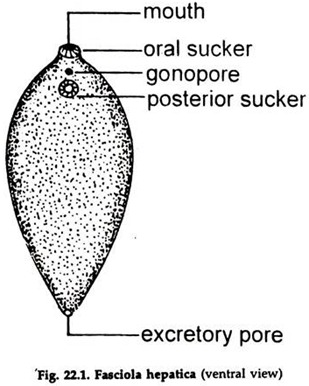
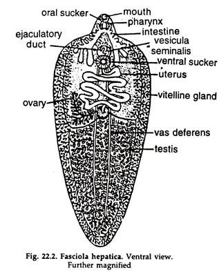
a. Body soft, flattened, leaf-like with a triangular head lobe (Figs. 22.1 & 22.2). It is brown to pale-grey in colour and measures 2.15-3.0 cm x 1.2-1.5 cm. The body is covered with a cuticle, the greater portion of which bears minute spines.
b. The mouth is anterior and terminal, surrounded by the oral sucker.
c. The posterior sucker is ventral and behind the mouth.
d. The genital aperture is placed in between the two suckers.
ADVERTISEMENTS:
e. The excretory pore is median and posterior terminal.
2. Body Wall of Fasciola (Fig. 22.3):
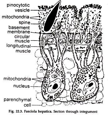
i. The outer surface is covered by a syncytium known as tegument.
ii. In light microscopy the tegument appears as a non-cellular homogeneous layer of about 7-16 µm thick.
ADVERTISEMENTS:
iii. Electron microscopic study reveals, it has two distinct zones. The outer zone consists of cytoplasmic syncytium; embedded in it are mitochondria, endoplasmic reticulum, various types of vacuoles, glycogen granules and other inclusions.
iv. The outer surface is thrown into numerous microvilli (folds). Some tegumentary spines are also present which are over-layed by the superficial plasma membrane.
v. The outer syncytial zone is connected by cytoplasmic bridges to inner nucleated bodies, known as cytons or perikarya. Collectively the cytons form the inner integumentary zone.
vi. In between the two layers lies a thin layer of connective tissue fibres known as basal lamella. Beneath this are a series of circular muscles under which lies the longitudinal muscle bands.
3. Digestive System of Fasciola:
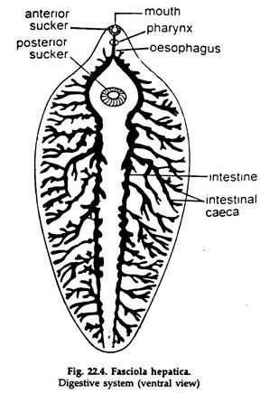
i. Digestive canal (Fig. 22.4) begins from a ventrally situated mouth, surrounded by the oral sucker. The mouth leads into a small funnel-shaped buccal cavity. The latter is in connected with pharynx.
ii. The pharynx is a highly muscular, rounded, thick-walled structure provided with pharyngeal gland.
ADVERTISEMENTS:
iii. The pharynx leads into a short, narrow oesophagus.
iv. The oesophagus connects the intestine, which immediately divides into right and left caecum; running up to the posterior end and terminates blindly. Each caecum on its side gives out numerous blind branches extending almost to all parts of the body.
v. Liver fluke feeds on tissue elements and exudates including bile, blood, and lymph.
vi. Fasciola lacks circulatory system, the ramification of digestive canal helps in distributing nutrients to different parts at the body.
vii. Digestion is mostly extracellular. Intracellular digestion also occurs.
viii. Undigested wastes are egested through the mouth. The alimentary canal is really a glorified coelenteron.
4. Excretory System of Fasciola:
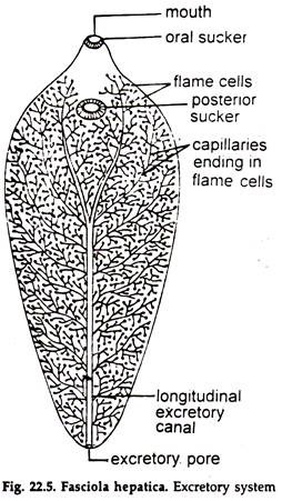
a. The excretory system is protonephric type. A single median longitudinal excretory duct opens posteriorly by a single excretory pore (Fig. 22.5).
b. Anteriorly, the excretory duct branches into four trunks, two dorsal and two ventral, each of which divides repeatedly to form smaller vessels. These smaller vessels branch repeatedly ending into flame cells.
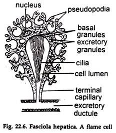
c. A flame cell (Fig. 22.6) is a modified mesenchyme cell.
i. It is of irregular shape, with granular cytoplasm and a nucleus.
ii. A bundle of flagella arises near the nucleus.
iii. The flagella are enclosed in a funnel, shaped space, formed by the end of a capillary, coming from the excretory duct and flicker constantly like a flame.
iv. The flame cells maintain a constant hydrostatic pressure by which wastes are driven into the excretory duct.
v. The cells are scattered in the parenchyma in a definite pattern and remove metabolic wastes from nearby tissues.
5. Nervous System and Sense Organs of Fasciola:
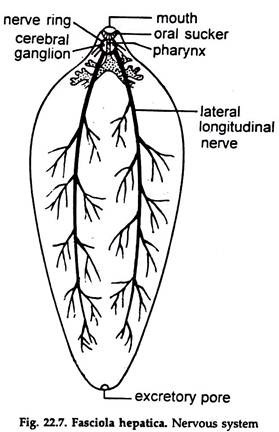
a. The nervous system (Fig. 22.7) consists of a cerebral ring encircling the oesophagus.
b. A pair of lateral cerebral and a ventral ganglia are present over the cerebral ring.
c. A number of slender nerves arise from the ganglia, run forward and innervate the head lobe.
d. Three pairs of longitudinal nerve cords— dorsal, ventral and lateral—are given out from these ganglia, which run backward.
e. The lateral pairs are long and stout and run posteriorly as lateral nerves, sending branches in their way.
f. Special sense organs are absent.
6. Reproductive System of Fasciola:
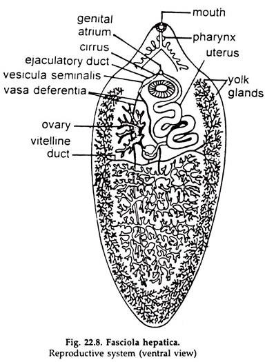
Fasciola is hermaphrodite and bears organs of both the sexes (Fig. 22.8). The common genital aperture is situated on the mid-ventral line, just anterior to the ventral sucker.
i. Male Reproductive Organs:
1. The testes are a pair, occupying the major part of the middle region of the body, one behind the other.
2. Each testis is a much branched-tubule, from the anterior end of which runs forward a narrow vas deferens.
3. Anteriorly, the two vasa deferentia join to form a median vesicula seminis, from which nans an ejaculatory duct, opening at the tip of the cirrus or penis, which, in turn, opens in the genital atrium.
ii. Female Reproductive Organs:
1. Ovary or the germarium is single, consists of much-branched tubules and located in the right hand side, in front of the testes.
2. The main oviduct is formed by the joining of the tubules.
3. Numerous rounded follicles, constituting the vitelline or yolk glands occupy considerable zone in the lateral regions of the body. They are connected with the vitelline ducts.
4. On each side, two large ducts, anterior and posterior, unite to form a single main lateral duct and there are two lateral ducts— right and left.
5. The lateral ducts run inward, nearly transversely and open into a small sac, the yolk reservoir.
6. Arising from the reservoir, a short, median vitelline duct runs forward to open into the oviduct.
7. Groups of unicellular shell glands or accessory female glands, opening by small ducts into the oviduct, are present around the junction.
8. The lumen of the oviduct at the region yolk is termed ootype.
9. The uterus is a wide, median, convoluted tube, formed by the union of the oviduct and the median vitelline duct, opens in the genital atrium, common to the external apertures of both the male and female ducts, near the base of the cirrus.
10. A canal, the Laurer’s canal, runs from the junction of the oviduct and median vitelline duct to open externally on the dorsal surface.
7. Fertilization and Development in Fasciola:
a. The eggs are large ovoidal structures with brown colour due to the presence of bile pigment. On the average, they measure, 40- 80 µm (microns). The eggs are fertilized in the uterus and self- or cross fertilization may occur.
b. The zygote receives yolk and a chitinoid covering in the ootype, remains in the uterus for a short period and is discharged. Passing down the bile duct, the zygote reaches the intestine of the host and passes to the exterior with the faeces.
c. Active development starts and after 2-3 weeks the miracidium larva hatches out of the egg.
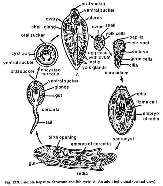
8. Life History of Fasciola (Fig. 22.9):
Miracidium Larva:
a. The body is conical with a triangular head lobe and is covered with vibratile cilia.
b. A pair of crossed eye spots are present anteriorly.
c. An imperfectly developed intestine and a pair of flame cells opening to the exterior are present.
d. The rest of the interior is filled up with a mass of germ cells.
4. The miracidium swims actively in water or moves on damp herbage and can survive only if it reaches an amphibian snail, the other host, approximately within eight hours-time.
5. The embryo bores into the snail and comes to the pulmonary sac or other organs.
6. The ciliated ectoderm is lost, it grows to an elongated sac and forms the sporocyst.
Sporocyst:
a. The body wall is formed by a single layer of cells.
b. The flame cells and remnants of eye spots are present.
c. The internal cavity contains germ cells.
7. The germ cells of the sporocyst behave like parthenogenetic ova. Each cell divides to produce blastula, gastrula and finally a form of larva, the redia.
8. Five to eight rediae are usually formed in each sporocyst.
Redia:
a. The body is cylindrical, with a circular ridge near the anterior end and a pair of short processes near the posterior end.
b. The mouth leads into pharynx and a sac-like intestine is present.
c. A system of excretory vessel is present.
d. Germ cells are present in the internal cavity.
e. A birth opening is situated anteriorly near the circular edge.
9. From the germ cells in the redia, 14-20 cercariae develop in each redia. Cercariae are produced if the season is summer but redia gives rise to a fresh generation of 8-12 rediae if the season is winter.
10. The cercaria escapes through the birth opening of the redia.
Cercaria:
a. The body is oval with a long tail.
b. The anterior and posterior suckers, the mouth, pharynx and a bifid intestine are present.
c. The gonads, glands, etc. begin to appear.
11. The cercaria forces its way out of the organ of the snail and reaches pulmonary sac, from where it escapes outside. It loses its tail, becomes encysted and remains attached to blades of grass or other herbage.
12. The encysted cercaria known as meta-cercaria, is taken by the herbivorous host with the grass and occasionally by man with vegetables. The metacercaria excysts in the duodenum and the young fluke, on escape, may reach the liver through bile ducts or hepatic portal vein, and grows rapidly to reach the adult stage. The eggs come out with the faeces about 3 to 4 months (incubation period) after infection.
9. Effect of Parasitism on Fasciola:
On Host:
The attack of liver fluke causes ‘liver-rot’, which is disastrous to the host and death has been recorded in most cases of liver-rot. Jaundice and adenomata have also been reported.
On Parasite:
Due to parasitic life, considerable degeneration of the vegetative organs has taken place in Fasciola. On the other hand, the reproductive organs are more developed.
A single fluke may produce about 50,000 eggs. Twice in its life cycle, the embryos are exposed to the environment and the cycle— which is already full of risks—becomes more risky. To compensate the huge loss during its perilous journey from host to host further multiplication by asexual means has appeared, in addition to the already accentuated rate of multiplication.
