In this article we will discuss about:- 1. Structure of Taenia Solium 2. Body Wall of Taenia Solium 3. Excretory and Osmoregulatory Systems 4. Nervous System and Sense Organs 5. Reproductive System 6. Fertilization and Development 7. Life History.
Contents:
- Structure of Taenia Solium
- Body Wall of Taenia Solium
- Excretory and Osmoregulatory Systems of Taenia Solium
- Nervous System and Sense Organs of Taenia Solium
- Reproductive System of Taenia Solium
- Fertilization and Development in Taenia Solium
- Life History of Taenia Solium
1. Structure of Taenia Solium:
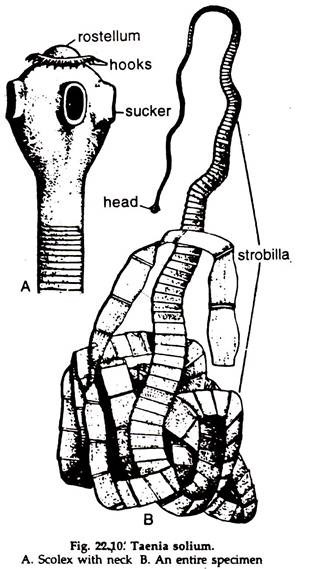
i. Body ribbon-like, 2-3 metres long and consists of about 800-900 segments, known as proglottids (Fig. 22.10).
ii. The head or scolex is small but distinct, pear-shaped and bears a conical, retractile elevation, the rostellum at the tip.
ADVERTISEMENTS:
iii. The rostellum bears 22 to 33 hooks, small and large, alternately placed and arranged in two rows.
iv. Four cup-shaped suckers are present on the sides of the head at about the middle. The hooks and rostellum anchor the parasite to the intestine of the host.
v. A small, un-segmented, narrow neck is present behind the head.
ADVERTISEMENTS:
vi. The rest of the body is ribbon-like segmented and called strobilla. The proglottids progressively enlarge in size posteriorly.
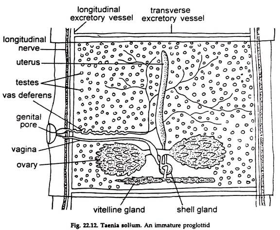
a. A proglottid at about the middle of the strobilla is roughly rectangular in outline (Fig. 22.12). It is thick and muscular.
b. A longitudinal nerve and a pair of longitudinal excretory canals are present in each lateral side of a proglottid.
c. A transverse excretory canal connects a pair of longitudinal canals at the posterior end of the proglottid.
ADVERTISEMENTS:
d. Numerous testes, their efferent ducts, a vas deferens, a pair of ovaries, two oviducts, a median oviduct, a single vitelline gland, an uterus, shell glands and an ootype are present in each proglottid.
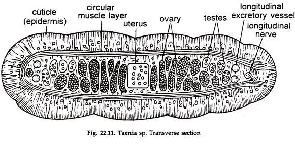
2. Body Wall of Taenia Solium (Fig. 22.11):
a. From the integument, the outer limiting membrane project numerous specialized microvilli, called microtriches (singular, microtrix).
b. Each microtrix possesses an electron- dense apical tip separated from the more basal region by a multi-laminar plate. The entire surface of the tegument is covered by a layer of carbohydrate-containing macro- molecules known as glycocalyx.
c. The cytoplasmic part of the microtriches are syncytial which are continuous with the external level of the tegument.
d. The external and internal levels of the tegument are joined by cytoplasmic bridges.
e. The external level of tegument contains numerous electron dense organelles like mitochondria, Golgi bodies, ribosomes, and endoplasmic reticulum. In addition, a number of vacuoles, vesicles and rhabdiform organelles, and glycogen granules occur.
ADVERTISEMENTS:
f. The internal levels of the tegument consists of discrete cells known as cytons or perikarya.
g. The cyton, a large cell, includes a prominent nucleus, mitochondria, Golgi bodies, endoplasmic reticulum, ribosomes and some vesicles.
h. The basal lamina, a layer of connective tissue, immediately beneath the external level of the tegument.
i. Beneath the basal lamina is the muscle layers. Musculatures are arranged in two layers. The outer layer is circularly or transversely arranged and the inner layer is longitudinally oriented.
j. A thin layer of neuromuscular cells lies underneath the muscles of the body wall; some cytoplasmic processes of these cells join with other similar processes of the cells while some other processes join with fine nerve fibres.
k. The space enclosed by the body wall except for that occupied by the reproductive organs, muscle fibres, nervous tissue and osmoregulatory structures, is filled with a spongy tissue called parenchyma. The parenchymal cells serve as sites for synthesis and storage of glycogen.
3. Excretory and Osmoregulatory Systems of Taenia Solium:
The excretory organs not only excrete metabolic wastes but also maintain hydrostatic pressure in the body. Thus, the excretory system also helps to maintain water balance of the body. In Taenia, the excretory system is protonephric type as in trematods and consists of four longitudinal canals, transverse canals and flame cells. The flame cells resemble those of the liver fluke.
Excretory Canals:
i. Four main collecting canals (ducts) extend the entire length of the strobilla. The two ventral canals are ventrolaterally located and the two dorsal canals are dorsolateral. All the canals are situated in the peripheral zone of medullary parenchyma.
ii. A single transverse canal connects the two ventral canals at the posterior end of each proglottid.
iii. The ventral canals carry fluid away from scolex, the dorsal canals towards it.
iv. Within the scolex, the four longitudinal canals may be joined by a network of canals or by a single ring canal or the dorsal and ventral canals of each side may be joined by a simple connection with no apparent exchange between the two sides.
v. Along the length of the ventral canals, a series of secondary tubules give rise to tertiary tubules.
vi. At the free end of the terminal tubules, flame cells are generally arranged in groups of fours.
4. Nervous System and Sense Organs of Taenia Solium:
The nervous system consists of a ‘brain’ in the scolex and nerves extending both anteriorly and posteriorly.
a. The brain is more or less rectangular and lies in the scolex.
b. The anterior long side of the rectangle is the anterior commissure, the posterior long side is the posterior commissure.
c. The rectangle itself is the cephalic ganglia.
d. Two long major trunks extend posteriorly along the entire length of the strobilla.
e. Two shorter nerve trunks proceed anteriorly to innervate the tissues anterior to the brain’.
f. Nerves from the trunks extend to the muscles, tegument, reproductive and other systems.
g. Sense organs are lacking.
5. Reproductive System of Taenia Solium:
The Taenia is hermaphrodite (Fig. 22.12). The reproductive organs first appear at about 200th segment, the male part appearing first. The reproductive organs attain maturity at about 400th segment and the posterior segments are filled up with a greatly increased uterus, heavily laden with developing embryos.
Male Reproductive Organs:
i. The testes are numerous, present throughout the proglottid and each testis is connected with an efferent duct The efferent ducts unite to form larger ducts and finally by joining the larger ducts a vas deferens is formed.
ii. The convoluted vas deferens runs to the lateral margin of the proglottid, becomes narrow and passing through a protrusible process, the cirrus opens in the genital atrium. At the base, the cirrus is enclosed in a muscular cirrus sac.
Female Reproductive Organs:
i. The ovaries or germaria are paired, bilobed and located at the posterior region of the proglottid.
ii. Numerous branching tubules constituting the ovary, join to form an oviduct. The two oviducts join medially to form a common oviduct.
iii. The vitelline or yolk gland is single, lobulated and situated at the posterior border of the proglottid, opening in the median oviduct through a yolk duct.
iv. Numerous shell glands are located around the yolk duct and open through their ducts into the oviduct at its junction with the yolk duct.
v. The part of the oviduct receiving the shell gland ducts and the yolk duct is termed ootype. Anteriorly, it is continuous with a median, elongated, blind uterus.
vi. The female genital pore located in the genital atrium, leads to a narrow passage, the vagina, which runs backward to the middle of the segment and dilates at the end to form receptaculum seminis, which is usually filled with sperms.
vii. The fertilizing or spermatic duct, runs from this duct to join, the oviduct.
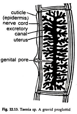
viii. The simple, cylindrical uterus begins to branch from at about 600th segment. The other reproductive organs degenerate and the gravid uterus, heavily laden with developing embryos look like a longitudinal stem with 5-10 branches (Fig. 22.13) and occupies practically whole of the segment in the posterior part of the strobilla.
6. Fertilization and Development in Taenia Solium:
1. Eggs are fertilized in ‘he oviduct and self- or cross-fertilization may take place.
2. The zygote becomes surrounded by yolk and a chitinoid egg shell, and passes to the uterus.
3. The ripe proglottids loaded with developing embryos get detached in chains of 5 or 6, and pass to the exterior with the faeces of the host.
4. The proglottids are eaten up by pig muscles of the segments get digested and six-hooked or hexacanth embryos are liberated.
7. Life History of Taenia Solium:
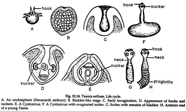
Hexacanth embryo or onchosphere (Fig. 22.14):
a. The body is rounded and enclosed in two membranes.
b. Six curved chitinoid hooks appear at one end and the hexacanth embryo is formed. The embryo, along with the membranes, is termed onchosphere.
c. The embryos bore the wall of the gut of the host with the hooks and enter blood vessels. From there they enter the general circulation, usually through the portal vein and reach the following organs in succession → liver → right side of the heart → lungs → left side of the heart → the systemic circulation. The embryo is filtered out and finally enters the muscular tissue.
d. Here the embryo increases greatly in size, having a large cavity filled up with watery fluid and assumes a bladder-like structure.
e. A hollow invagination or ingrowth takes place at one point of the bladder and on the inner surface of this invagination develop hooks and suckers, characteristic of the head of adult Taenia.
f. The hollow ingrowth becomes everted and the suckers and hooks come close to the surface.
g. The embryo now looks like a bladder with a head and neck of Taenia lying within it. This is known as bladder worm or cysticercus stage. It is rich in salt and albuminous material and when the pig’s flesh is infected with the cysticercus cellulose, it is known as measly pork.
h. If a portion of measly pork is eaten by man, the bladder is dissolved by the gastric juice. The gastric juice causes the albuminous material to swell up and forces the fluid into the cavity, thereby the head comes out through the pore. The scolex attaches itself to the wall of the intestine by hooks and suckers and develops the series of proglottids to reach the adult stage.
i. The worm gains sexual maturity by 2-3 months’ time, and survives for 25 years or more.
