The following points highlight the top seven types of excretory organs in invertebrates. The types are: 1. Contractile Vacuoles in Paramoecium 2. Excretory System of Planeria (With Reference to Flame Cells) 3. Excretion in Taenia Solium 4. Excretion in Earthworm with Reference to Nephridia 5. Excretion in Cockroach with reference to Malpighian Tubules and Others.
Excretory Organs in Invertebrates:
- Contractile Vacuoles in Paramoecium
- Excretory System of Planeria (With Reference to Flame Cells)
- Excretion in Taenia Solium
- Excretion in Earthworm with Reference to Nephridia
- Excretion in Cockroach with reference to Malpighian Tubules
- Excretion in Prawn with Reference to Green Gland
- Excretion in Bivalve (Lamellidense) with Reference to Organ of Bojanus
Excretory Organs in Invertebrates: Type # 1.
Contractile Vacuoles in Paramoecium:
ADVERTISEMENTS:
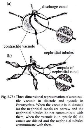
In Paramoecium, two large contractile vacuoles are present almost at the fixed position one near each end of the cell. One vacuole is located at the anterior part and the other at the posterior part of the body. Each vacuole consists of a large central vesicle into which opens a number of radiating or inhalant canals.
The radiating canal is divisible into three parts:
(i) Outer-terminal part,
(ii) Middle-ampulla and
ADVERTISEMENTS:
(iii) Inner-injector canal.
Electron microscope studies reveal complex systems of tubules which are connected with the endoplasmic reticulum and empty into the contractile vacuole through a clearly defined system of canals. These vacuoles are always associated with the innermost region of the cytoplasm and empty to the outside through one or two permanent pores that penetrate the pellicle.
Water Elimination:
The water separation takes place over large areas of intracytoplasmic surface and involves the formation of minute droplets along the tiny tubules. Cation pump reduces the tonicity of the fluid of the droplets.
ADVERTISEMENTS:
Soon a wave of contraction in the coacervate cytoplasmic gel surrounding the ampullae is generated. Fluid is pumped into the major central vacuole by these cytoplasmic movements that press around the ampullae from distal to proximal ends (Fig. 2.73).
Discharge of Vacuolar Fluids:
The movements of the endoplasmic gel generate the major contractile force. This force pushes the vesicle against the plasma lemma, bulging it until it ruptures. Fluid then rushes out, the vesicle collapses and inverts into itself. The contractile fibres on the central vesicle contribute very little in water expulsion.
Flame Cells:
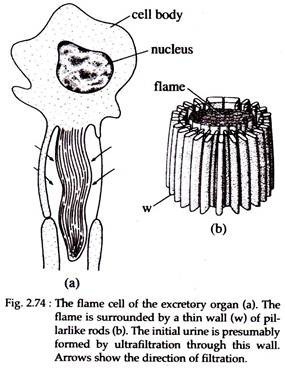
This is a type of protonephridia (Fig. 2.74). Protonephridia occur mainly in animals that lack a true body cavity (coelom). The closed ends of protonephridia terminate in enlarged bulb-like structures, each with a hollow lumen and one or several long cilia inside. If there is a single cilium, the terminal cell is called a solenocyte; if there are numerous cilia projecting into a lumen, the structure is called a flame cell.
The tuft of cilia when undulates, resembles the flickering flame of a candle. Functionally both these cells are same. Each cell is provided with a large nucleus. The cytoplasm is granular. At the centre of the cell a large cavity bears a bundle of densely packed cilia.
Each cilium originates from a basal granule. The flame is surrounded by a thin wall of pillar-like rods. The cell membrane has extended pseudopodia like structure within the inter-cellular space of the body to increase the absorption surface.
Function:
ADVERTISEMENTS:
Flame cells function on the basis of filtration and reabsorption. The water from intercellular spaces are collected by the extensions of the plasma lemma. The collected water is ultra-filtered through the thin wall of pillar-like rods. The filtered water then moves through the neck of the cavity by the flickering movement of the cilia.
It is also suggested that the constant beating of cilia could produce sufficient negative pressure to cause ultrafiltration. During movement of water through the neck region of the cavity, some of it are reabsorbed. Ultimately hypertonic urine is discharged to the exterior.
Excretory Organs in Invertebrates: Type #
2.
Excretory System of Planeria (With Reference to Flame Cells):
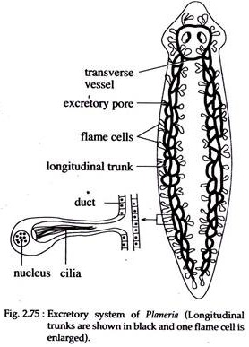
Excretory system or the system of water vessels in Planeria, consists of two pairs of longitudinal trunks, right and left, which rim through the whole body length (Fig. 2.75). Each trunk is much coiled and is connected with each other by transverse vessels in the anterior region and opens to the outside by means of several pairs of pores which are situated on the dorsal surface. The lumen of the vessels are lined by ciliated cells.
The trunks give off numerous branches within the body. From these branches extremely fine capillary vessels emerge. Many of these capillary vessels bear flame cells or flame bulbs or paranephridia at their tips. The flame bulb is a large cell bearing many cytoplasmic projections.
The nucleus is displaced to one side of the cell and the cytoplasm bears secretory droplets and is hollowed out to form a large central cavity which is continuous with that of the capillary vessels. A bunch of cilia hangs down into the cavity of the cytoplasm.
The excretory products in fluid state enters from the parenchyma cells by diffusion. The cilia within the cavity of the cytoplasm beat continuously and keep the fluid circulating. It is also believed that the flame bulb helps in the elimination of excess of water, i.e. the help in osmoregulation.
Excretory Organs in Invertebrates: Type #
3.
Excretion in Taenia Solium:
Excretory System:
There are two pairs of main longitudinal excretory trunks in Taenia solium. Each pair runs along with the lateral margins of the strobila. The paired state of the longitudinal trunks is well recognised at the anterior part, of the strobila and in the posterior part one from each of the pairs becomes lost. The two pairs of the longitudinal trunks are connected with each other in the head region by a ring-like vessel.
Similar connections by straight transverse vessels are seen in the posterior region of each proglottid. In the last and penultimate proglottid the longitudinal trunks open into a pulsatile caudal vesicle which opens to the outside by a single median aperture.
When the last proglottid is cast off at maturity, the longitudinal trunks open to the exterior separately and independently. The main trunks give of branches from which numerous fine canaliculated arise, each terminating in a flame cell. The structure of the flame cell is similar to that of Planeria.
Excretory Organs in Invertebrates: Type #
4.
Excretion in Earthworm with Reference to Nephridia:
Excretory System:
Excretory organs are the nephridia. Nephridia occur in all segments excepting the first three and the last segments. The nephridia are small and coiled tubular structures and occur in huge numbers. In the body of earthworm three kinds of nephridia occur—septal, integumentary and pharyngeal. Structurally, these nephridia show basic similarities its classification is based on its position in the body.
(a) Septal Nephridia:
Septal nephridia remain attached to the two faces of the septum. They occur from 15th segment backward. That means in the first fourteen segments they are absent. Each septum bears 40- 50 nephridia in average in its anterior and posterior face. Thus, in each segment there are about 80-100 nephridia.
Structure of Septal Nephridium:
A typical septal nephridium (Fig. 2.76) consists of a main body formed by a straight lobe and a long narrow, spirally twisted loop, a funnel-like nephrostome connected to the main body by a short neck and a terminal nephridial duct. All the nephridial structures remain restricted to the same segment.
The nephrostome or funnel is a rounded structure. The mouth of the funnel which communicates with the coelom is provided, with a large upper lip and a small lower lip. The lips are ciliated. A narrow ciliated tube runs from the funnel into the body of the nephridium and takes several turns inside it.
The main body of the nephridium is made up of a main lobe and a spirally twisted loop. The loop is twice as long as the main lobe and consists of a proximal limb and a distal limb twisted round each other.
The straight lobe is continued as the distal limb of the twisted loop and the proximal limb receives the ciliated tubule from the nephrostome and also gives off the terminal duct which opens at the nephridiopore. The straight lobe bears four parallel tubules, the proximal and distal ones bear three tubules each and in the apical part there are two tubules.
Terminal ducts of the septal nephridium open into a septal excretory canal which runs parallel and internal to commissural vessels. There are a two of septal excretory canals situated one on each side of the septum.
The two septal excretory canals open into a pair of supra-intestinal excretory ducts which run on the mid-dorsal line, side by side, from 15th segment to the posterior end. The supra- intestinal excretory ducts open into the lumen of the intestine by a small duct at the level of each inter-segmental septum.
(b) Integumentary Nephridia:
They are smaller in size than the septal nephridia. These ‘V’-shaped structures occur on the inner surface of the integument in all segments excepting the first two.
They occur 200-250 in each segment but in the 14th, 15th and 16th segments the number of nephridia is much more. Structurally they resemble septal nephridia but lack the nephrostome. They open independently to the outside by nephridiopores on the outer surface of the body wall.
(c) Pharyngeal Nephridia:
They are as large as the septal nephridia and occur in three pair, as bunches or tufts, in the 4th, 5th and 6th segments on either side of pharynx and oesophagus. Nephrostomes are also absent in the pharyngeal nephridia. In each bunch the terminal ducts of the nephridia join together to form a slender duct.
The slender ducts again unite in each segment and form a thick walled duct which opens into the alimentary tube. Thus there are three pairs of ducts, one pair each in the 4th, 5th and 6th segments. Some workers mention that the pharyngeal nephridia have digestive function or, in other words, they aid in digestion and hence they are sometimes referred to as “peptic nephridia”.
The septal and pharyngeal nephridia open into the alimentary canal and are called enteronephric while the integumentary nephridia open to the outside directly and are called exo-nephric. The enteronephric system helps in the conservation of water in the body because the water present in the excretory product is again reabsorbed in the intestine.
Process of Excretion through Nephridia:
The funnel shaped nephrostome collects the fluid from coelom. The fluid then passes down through the extensively looped duct of the nephridia. The fluid, at the time of entering the nephridium is isotonic, but during its travel through the nephridium, salt is withdrawn and a dilute urine is discharged.
Therefore, the nephridium is functioning as a filtration – reabsorption organ, in which an initial fluid is formed by ultrafiltration and subsequently modified by reabsorption.
Other Structures Associated with Excretion in Earthworm:
Chloragogen Cells:
These cells (Fig. 1.96A) form chloragogenous tissue which lie close against the wall of intestine. These are specialized derivatives of the coelomic epithelium. Chloragogen cells store fat and carbohydrate and they are also the main site of deamination.
Ammonia and urea pass from these cells into the coelomic fluid and are swept through the nephridial funnel into the tubule. The waste material, including minerals, which are accumulated in these cells are carried out by amoebocytes. These materials are either deposited or passed through the epidermis.
Excretory Organs in Invertebrates: Type #
5.
Excretion in Cockroach with Reference to Malpighian Tubules:
Excretory System in Cockroach:
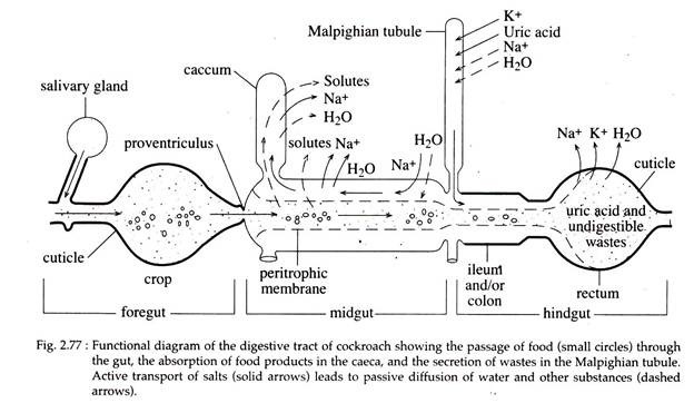
Near the junction of the mid gut and hind gut, there are numerous (70-120) threadlike, blind tubes, called Malpighian tubules, which remain arranged in six bundles (Fig. 2.41). Each bundle contains 15-20 tubules.
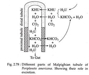
The proximal end of each tubule opens within the lumen of the gut and the distal blind end extends within the haemocoel (Fig. 2.78). The tubules are of two kinds—one kind takes darker silver nitrate stain than the other. The metabolic wastes which are collected from the haemocoelomic fluid are finally drained within the cavity of the hind gut.
Process of Excretion by Malpighian Tubules:
Potassium is actively secreted into the lumen of the tubule from the coelomic fluid. Water, along with urea and Na+ follow passively, due to osmotic forces (Figs. 2.77 & 2.78). Therefore, a potassium-rich fluid is formed in the tubule.
However, this fluid is isotonic to blood, but has a very different composition. The fluid then enters into the hind gut. Here, the solutes and much of the water are reabsorbed and uric acid is precipitated. The remains of the fluid in the rectum is eventually expelled out as a mixture of urine and faeces.
Accessory Excretory Structures of Cockroach:
(a) Nephrocyte or Pericardial Cells:
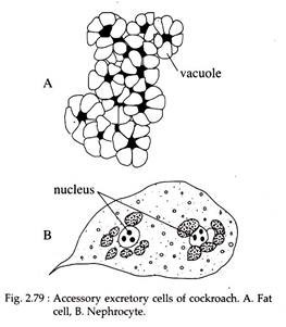
These are aggregated cells typically located on the heart. Sometimes may exist singly. Large cells are found with numerous vacuoles engorged with excretory granules (Fig. 2.79B). It is reported that they absorb excretory materials from haemolymph and store them in the cytoplasm.
(b) Rectal Glands:
The papillae of the internal wall of the rectum, is composed of a special type of glandular cells called rectal glands. These glands assist in reabsorption of water and minerals from rectum and therefore, help in osmoregulation.
(c) Fat Cells:
The fat mass of the body cavity is composed of fat cells (Fig. 2.79A). These cells are primarily fat storing cells. But along with fat they also store urate in the cytoplasm.
(d) Moulting or Amoeboid Cells:
During nymphal stages some amoeboid cells store excretory material and remain attached with the cuticle. When the nymph changes its stage (metamorphoses), these amoeboid cells are removed along with the cuticle.
Excretory Organs in Invertebrates: Type #
6.
Excretion in Prawn with Reference to Green Gland:
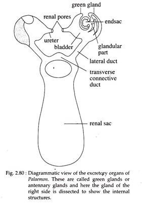
Excretory organs of prawn are known as green glands or antennary glands. These are paired (Fig. 2.80) white organs which remain within the coxa of each second antenna.
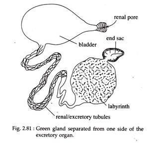
Each organ consists of following parts (Fig. 2.81):
(a) End Sac:
This small bean-shaped structure contains blood lacunae. Its wall is two- layered. The inner layer of epithelial cells having excretory function and the outer thick connective tissue layer has minute lacunae. Radially arranged partitions called septa, project from the wall within the central cavity.
(b) Labyrinth:
It is present outside the end sac and contains many narrow, branched and coiled excretory tubules. Each tubule communicates with the end sac by a single opening but opens within the bladder through several apertures. A single epithelial cell layer having excretory function, lines each tubule.
(c) Renal Tubule/Excretory Tubule:
It is a coiled and thin walled tubule emerging out of the labyrinth and ends in a bladder,
(d) Bladder:
It is a thin-walled sac with an epithelial lining. It communicates with the exterior through a small ureter.
(e) Excretory Opening:
It is present on the base of each second antenna. Both the green glands are connected with a- common large thin-walled transparent and centrally placed sac called the renal sac (Fig. 2.80). It is present between the cardiac stomach and the carapace and it communicates with the bladder of each green gland by a separate lateral duct. The two lateral ducts are interconnected by a transverse connective duct.
End sac and the labyrinth are the two regions responsible for extracting urine from the blood. The excretory product includes ammonia (excreted by end sac), uric acid, other- nitrogenous compounds and excess water (excreted by other parts). The urine remains temporarily stored within the bladder and is periodically expelled out through the renal apertures.
In addition to the green glands, gills and integumental coverings are also responsible for excretion. The exoskeleton at the time of its periodic replacement carries a large quantity of excretory products.
Process of Excretion:
The saccules collect fluid by filtration with the help of podocytes present in its wall. The greatly folded, glandular labyrinth is the site of reabsorption. The saccules filters some specific ions, mainly Mg++. Therefore, it is believed that green gland controls internal fluid pressure. For this purpose it reabsorbs Mg through labyrinth. A copious amount of urine may be excreted, depending on internal and external conditions.
Although green gland controls internal fluid pressure through the regulation of specific ions, i.e. Mg++, they do not play a major role in osmotic regulation. Lastly, although, these are called excretory organs, they never excrete nitrogenous wastes. Most nitrogenous wastes (NH3) are diffused through gills or from the body surface where the exoskeleton is thin.
Excretory Organs in Invertebrates: Type #
7.
Excretion in Bivalve (Lamellidense) with Reference to Organ of Bojanus:
Excretory System:
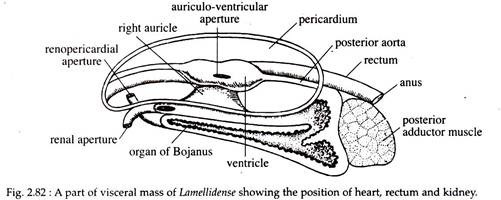
The excretory organs of Lamellidense comprise of a pair of kidneys called the organ of Bojanus. These are situated one on each side of the body below the pericardium. Each kidney consists of two parts, a glandular part and a thin-walled ciliated urinary bladder (Fig. 2.82).
The glandular part of the kidney opens into the pericardium by Reno-pericardial aperture and the bladder opens to the exterior by a minute opening called renal aperture (nephridiopore) situated between the visceral mass and the inner lamina.
In addition to kidneys, a large reddish-brown glandular mass known as Keber’s organ or pericardial gland is regarded to be excretory in function. It is situated in front of the pericardium and discharges the excretory products into the pericardial cavity.
Process of Excretion:
Urine originates as an ultra-filtrate from the heart into the pericardium. From the pericardium it passes through the glandular region of the kidney. Here the ionic composition of the urine changes—i.e., reabsorption of the minerals and water takes place.
The principal excretory products, ammonia and urea, comes out through renal aperture. In many bivalves, the renal organs play an important role in osmoregulation, a role that sometimes exceed that of excretion.
