In this article we will discuss about the Macrophagy in Hydra:- 1. Subject Matter of Hydra 2. Feeding in Hydra 3. Process 4. Digestion.
Subject Matter of Hydra:
Hydra is an animal under Phylum Cnidaria. It has no head end. The mouth (which serves also as the anus) is the single opening of the only internal cavity, called the coelenteron. At the oral end the mouth is situated on a cone like structure called the hypostome.
Around the base of the cone is a circle of about six tentacles. On the tentacles stinging cells are concentrated. Stinging cells called cnidae or nematocysts are used for food capture and defence. Nematocysts are unique to the phylum and a diagnostic feature of it.
Feeding in Hydra:
Hydra is carnivorous and feeds mainly on small crustaceans. For capturing the food, they possess some specialised cells.
ADVERTISEMENTS:
The structure and functions of these cells are described below:
Cnidocytes/Cnidoblast cells:
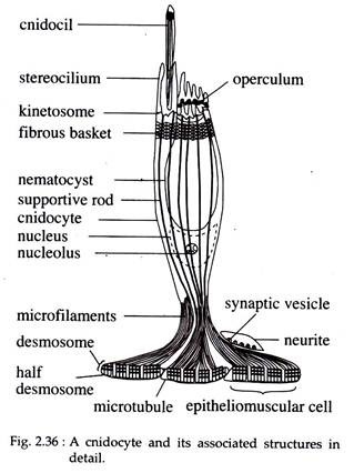
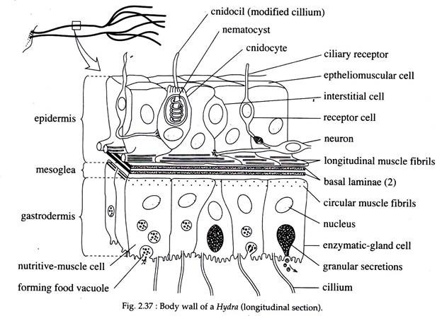
They are located throughout the epidermis of the tentacles and are lodged between or invaginated with epitheliomuscular cells of the epidermis (Fig. 2.37). A cnidocyte is a rounded or an ovoid cell with a basal nucleus.
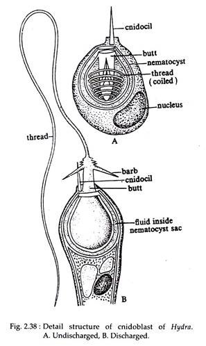
One end of the cell contains a short, stiff, bristle-like process called a cnidocil (Fig. 2.36 & 2.38). The cnidocil in electron microscope, reveals structural similarity with that of a cilium. It remains exposed to the surface.
ADVERTISEMENTS:
The interior of the cell is filled by a capsule containing a coiled, usually pleated tube. The outwardly directed end of the capsule is covered by an operculum or by lid-like flaps. The base is attached to the lateral extensions of one or more epitheliomuscular cells. Sometimes these cells may be associated with a neuron terminal.
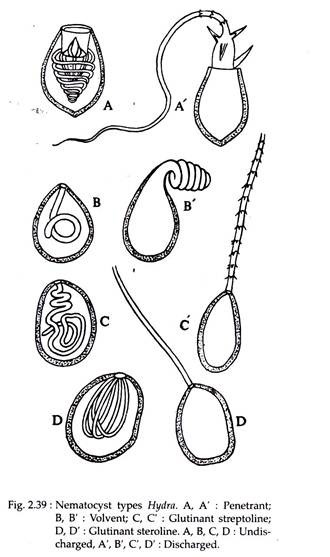
The commonest type of cnidocyte is the stinging nematocyst (Fig. 2.38). In addition to the structures of general cnidocytes, these cells possess a butt, stylet and spines. The capsular fluid is toxin and a mixture of protein and phenol. Hydra possess four types of nematocysts (Fig. 2.39).
(a) Penetrant or Stenoteles:
They have large capsule with a thick and large butt. Last row of spines are modified into stylet. It acts as an organ of offence and defence.
(b) Volvent or Desmoneme:
Smaller than penetrant and oval in shape. Small, thick tube that has no spines or butt. They coil round the prey and help in capturing food.
(c) Small Glutinant or Atrichous Isorhiza:
They are like volvent nematocyst, except the possession of a thread with terminal aperture. It helps to fix the tentacle on the substratum.
(d) Large Glutinant or Holotrichous Isorhiza:
The butt and the thread are provided with spines. The capsule is oval and large. It also helps in capturing the prey.
Process of Food Capture in Hydra:
ADVERTISEMENTS:
The whole process of food capture can be described under the following headings (Figs. 2.40a, b, c & d):
(a) Recognition of the Prey:
The cnidocil of the cnidocyte, at first recognises a prey. The chemical and mechanical stimuli from the prey are perceived by cnidocil. It then transfers the stimuli to the interior.
(b) Discharge of Nematocyst Thread:
After getting the stimulus from the cnidocil, a sudden release of calcium within the capsule occurs. Water rushes into the capsule, and the intra-capsular pressure rises. This pressure plus some intrinsic tension within the capsule structure itself, causes the operculum or apical flaps of the cnidocyte to open.
Then the thread event. The nerve cell connections spread the information to the nearby nematocysts to work accordingly.
(c) Paralyzing the Prey:
After ejection, the penetrant nematocyst pierces the body of the prey and injects toxin into it. This toxin paralyses the prey. During paralysis the prey is kept within the territory of the tentacle by other nematocysts.
(d) Transfer of Prey into the Mouth:
A fully paralyzed prey is entangled and brought near the mouth by the tentacles. The tentacles holding the prey contract and bend over the mouth and help to transfer the food into the mouth. The prey is then engulfed by the movements of hypostome and mouth. The peristaltic contractions of the body wall force the prey into the coelenteron. In the coelenteron digestion begins.
Digestion in Hydra:
Both extracellular and intracellular digestions are present in Hydra. In the coelenteron, extracellular digestion occurs. Intracellular digestion occurs within the cells of the coelenteron wall. In case of Hydra, both these digestions are enzymatic — i.e., chemical. Thus, there are specialized cells that secrete different components of chemical digestion.
(a) Cells Responsible for Extracellular Digestion:
(i) Enzymatic gland cells (Fig. 2.37) discharge proteolytic enzymes. These cells are present in between nutritive muscular cells of the coelenteron wall.
(ii) Mucus secreting cells (Fig. 2.37) are also present in between nutritive muscle cells. They secrete mucus to enhance the activity of the enzymes.
(b) Cells Responsible for Intracellular Digestion:
Nutritive Muscle Cells:
These (Fig. 2.37) are epithelial cells of the coelenteron lining i.e., gastro-dermal lining. There are two types of nutritive muscle cell. One with flagella at its luminal end, while the other bears pseudopodia. They are capable of engulfing the half-digested food. In some species of Hydra, a symbiotic algae (Zoo chlorella) are often seen in association with these types of cell.
Stages of Digestion:
After the entrance of the prey into the coelenteron, the enzymatic gland cells discharge proteolytic enzymes. The mucus from mucus cells cover the prey and the proteolytic enzymes start acting on the prey. Gradually the prey-tissues are reduced to a soupy broth. The beating of the flagella of the gastro-dermal cells ensures mixing.
After this initial extracellular phase, digestion continues intracellularly. The nutritive muscle cells engulf small fragments of tissues in food vacuoles. Within the food vacuoles digestion of carbohydrates, fats, and proteins continues. The food vacuoles undergo the acid and alkaline phases like that of protozoans.
After digestion, the products are distributed to other cells by diffusion. Un-digestible materials are ejected into the coelenteron by exocytosis. From coelenteron the ejected materials are thrown out through the mouth when the body contracts.


