
In this article we will discuss about the Role of Microfilaments in Amoeboid Movement.
At present it is known that motive force in Amoeba is generated by sliding interactions between actin and myosin filaments in a way comparable to that occurring in muscle cells.
Actin in Amoeba:
Actin is the major structural protein in the force producing event. This contractile protein has been identified in Amoeba proteus and Acanthamoeba. These actins exist as G-actin or globular monomers and F- actin or polymeric filaments of G-actin units. Like muscle actin, actin in amoeba can reversibly bind with heavy meromyosin to form arrow-head shaped complexes visible under electron microscope.
ADVERTISEMENTS:
Myosin in Amoeba:
It acts as mechanochemical transducer in the force-producing event. It has been identified in A. proteus and Acanthamoeba. The protein has a globular head and rod like body comparable to heavy meromyosin and light meromyosin of muscle myosin’s. It can reversibly bind to actin filaments.
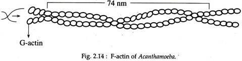
Micro-filamentous organisation of actin and myosin in amoeba cytoplasm: Thin microfilaments resembling muscle F-actin, have been identified in the cytoplasm of A. proteus and Acanthamoeba (Fig. 2.14).
ADVERTISEMENTS:
Electron microscopic study of the filaments following negative staining with uranyl acetate revealed that:
(i) each filament has an un-definite length but a width of 6-7 nm,
(ii) each filament has a double helical structure just like muscle F-actin.
Thick microfilaments, resembling the myosin filament of muscles have also been identified by electron microscopy from the cytoplasm of ATP- stimulated A. proteus. Each filament has a length of 500-700 nm and a width of 16-22 nm and each filament shows lateral projections like muscle-myosin filaments.