In this article we will discuss about:- 1. Habit and Habitat of Ascidia 2. External Structure of Ascidia 3. Body Wall 4. Coelom 5. Locomotion 6. Respiration 7. Circulatory System 8. Excretory System 9. Nervous System 10. Sense Organs 11. Reproductive System 12. Development and Life History.
Contents:
- Habit and Habitat of Ascidia
- External Structure of Ascidia
- Body Wall of Ascidia
- Coelom of Ascidia
- Locomotion of Ascidia
- Respiration of Ascidia
- Circulatory System of Ascidia
- Excretory System of Ascidia
- Nervous System of Ascidia
- Sense Organs of Ascidia
- Reproductive System of Ascidia
- Development and Life History of Ascidia
1. Habit and Habitat of Ascidia:
Ascidia is exclusively marine and occurs at all depths of the sea. It is sessile in adult stage, but the larva is a free-swimming form. The adults remain attached to the bottom of the sea or other substrata by its base. Ascidia is a microphagous animal, i.e., it feeds on microorganisms. The food materials are obtained by ciliary action.
2. External Structure of Ascidia:
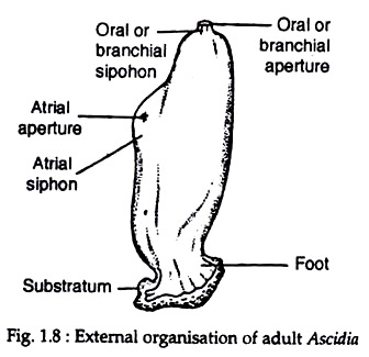
Ascidia, in adult stage, is a solitary animal having a bag-like body (Fig. 1.8). The body is covered by a tough and translucent tunic or test. Because of the presence of tunic, the urochordates are also known as tunicates. The tunic is composed of a carbohydrate, tunicin (chemically related to cellulose) and glycoprotein (20%).
ADVERTISEMENTS:
The most important cell-types are the large bladder-like cells. Numerous blood sinuses permeate the tunic. The free end of the body bears two apertures — one at the top called the oral funnel or oral siphon, containing the mouth, and the other is laterally placed and is called the atrial funnel or atrial siphon. The atriopore is situated in the middle of the atrial funnel. The oral funnel is usually eight-lobed, while the atrial funnel is six-lobed in Ascidia.
The atrial funnel is located at one side of the body. In living condition, current of water enters into the body through the oral funnel and goes out by the atrial funnel. If the animal is disturbed, the body wall contracts instantaneously, as a result, the water present inside the body is expelled out like jets.
Thus, the common name sea-squirt has been attributed to the specimen. The attached end of the body is called stolon or base.
3. Body Wall of Ascidia:
The test of Ascidia is referred as a cuticle. A number of branching tubes, each terminating in a tiny bulb-like dilatation, are present. Blood circulates through these tubes. Underneath the tunic, there lies the true body wall composed of living tissues called mantle.
ADVERTISEMENTS:
The mantle is covered by single-layered epidermis. The epidermis is composed of cuboidal or squamous cells. The mantle bears an underlying layer of connective tissues and muscle fibres which extend in various directions.
The longitudinal muscle fibres are numerous. Except at the regions of oral and atrial siphons the muscle fibres are oriented in an irregular network. They cross one another in all directions. Majority of the muscle fibres are either longitudinally or transversely disposed. Non-metameric organisation is a notable feature in Ascidia. This means the mesoderm remains un-segmented.
4. Coelom of Ascidia:
Due to extensive development of the atrium, the coelom is greatly reduced and exists in few doubtful derivatives, like pericardial cavity, gonadal cavities etc. Reduction of coelom is regarded to be a degenerative acquisition. The atrium is formed by involution of the body surface and it is not a coelom.
5. Locomotion of Ascidia:
ADVERTISEMENTS:
Ascidia, in adult stage, is a fixed form. So locomotion in true sense is absent. The movement is restricted to the contraction of the body by muscle fibres and the closure of the funnels. But the tadpole larva swims actively in water. The mechanism of swimming in the tadpole is exactly similar to that described in Branchiostoma.
6. Respiration in Ascidia:
The extensive ciliation in the pharyngeal cavity and the cilia bounding the stigmata set up a constant flow of water current passing inwards through the mouth. During the inflow of water current, the water (containing fresh oxygen in dissolved state) passes into the atrium via the stigmata.
This process is supplemented by the periodic muscular contractions of the body wall. From the atrium the water goes out to the exterior through the atriopore. The walls of the stigmata are highly vascular, where exchange of gases takes place. After oxidation, the resultant carbon di-oxide passes out along with the water from the atrium.
7. Circulatory System of Ascidia:
The circulatory system consists of blood, a heart, blood vessels and sinuses. The blood contains a colourless plasma and a few corpuscles. Ascidia lacks capillaries. As a result the blood and the tissue fluid become intermingled. The fluid contains lymphocytes, phagocytic macrophages, vacuolated compartment cells and many coloured and colourless cells.
Some ascidians possess vanadocytes (cells having green vanadium-containing pigment) in the plasma. The pigment is presumed to be respiratory pigment. Some of the corpuscles are phagocytic and the others contain pigments.
The heart is a fusiform sac-like structure which is surrounded by pericardium. The heart is situated below the pharynx. The heart communicates with a system of blood spaces called the haemocoel. Some of the larger spaces have an endothelial lining and thus form the blood vessels. Below the endostyle the heart gives a ventral or hypobranchial vessel to supply blood to the various parts of the pharynx.
From the opposite end of the heart another vessel called visceral vessel emerges out to supply blood to the viscera. The direction of flow of blood through the heart is periodically changed due to the reversal of peristalsis of heart muscles. Such a phenomenon is very rare and is not ordinarily observed in the circulatory system of any animal.
8. Excretory System of Ascidia:
There is no definite excretory organ in Ascidia. The nephrocytes present in the blood are found to accumulate excretory products in the cytoplasm. These cells contain particles of urates and xanthine. It is recorded that about 95% of the nitrogenous wastes are excreted in the form of ammonia. The neural gland also functions as the excretory organ.
ADVERTISEMENTS:
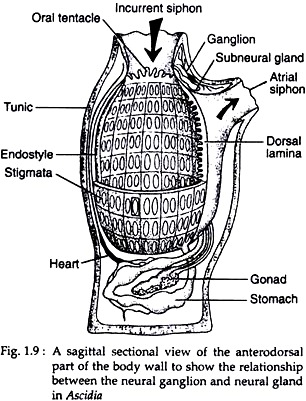
A peculiar hollow neural gland is found embedded in the ventral side of the nerve ganglion. The gland opens into the pharynx near the velum through a ciliated funnel (Fig. 1.9). This gland originates mainly from the larval ectoderm and partly from the pharynx. This double origin is quite significant. The gland is regarded to be a homologous structure, similar to the hypophysis of vertebrates.
9. Nervous System of Ascidia:
The entire nervous system is reduced to a single elongated solid nerve ganglion called cerebral ganglion or brain. It is situated dorsal to the neural gland and between the atrial and oral funnels. This ganglion gives off nerves, specially to the atrial and oral funnels and velum, and thus helps to regulate the flow of water current.
10. Sense Organs of Ascidia:
Sense organs, in the strict sense, are absent. The pigment spots or ocelli that are present in the adults around the funnels are regarded to be photo-receptors, in a slowly developed condition. But in a fully-grown tadpole larva this photoreceptive organ is highly developed.
11. Reproductive System of Ascidia:
Ascidia is a hermaphroditic animal. The gonads are sac-like structures situated in close association with the intestine. The gonads are mesodermal in origin. The ovary and testis possess elongated gonoducts. The gonoducts open into the atrium.
The sex cells are discharged into the atrium and from here are expelled to the exterior. Fertilisation is external. The sperms and eggs attain maturity at different times. Thus, self-fertilisation is prevented. Besides sexual reproduction, multiplication by budding (a process of asexual reproduction) also takes place. Ascidia is noted for its great potency of regeneration.
12. Development and Life History of Ascidia:
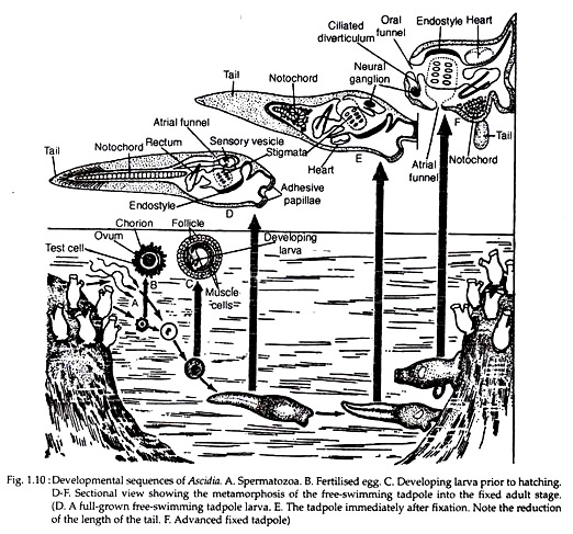
Development is indirect, i.e., development is accomplished through metamorphosis. The phenomenon of metamorphosis includes a series of changes which a larva has to undergo before attaining its adulthood. Ascidia furnishes the best example of metamorphosis in which a highly developed tadpole larva, in course of ontogenic processes, transforms into a sessile and degenerated adult (Fig. 1.10).
Pre-larval Stages:
The eggs are small and almost yolkless. Each egg is covered by some membranes which help it to float in water. Cleavage is holoblastic and nearly equal at the initial stages. A blastula with a few cells is produced. Gastrulation by invagination follows and the gastrula elongates. After about three days, a free-swimming tadpole larva emerges. The tadpole larva has all the characteristics of chordate organisation.
Larval Stages:
The larval form present in the life-history of Ascidia is called tadpole larva. It is very active at the beginning which, in course of metamorphosis, transforms into a sessile adult (Fig. 1.10D-F).
Structure of Early Tadpole Larva:
A fully grown tadpole larva is highly motile and does not take food from outside, i.e., it is a non-feeding form. It has an elongated body. The body is more or less oval in outline. It is distinctly divisible into two regions—the head and the tail (Fig. 1.10D). The whole of the body is covered by the tunic. The head is elliptical and has three adhesive papillae or chin warts.
Of the chin warts, one is mid- dorsally placed and the rest two are located ventrolaterally. The tail is laterally compressed and pointed terminally. It is provided with a caudal fin. The dorsal and ventral fins are continuous along the tail and are marked with striae. The striae are regarded to be precursors of the fin-rays in fishes.
The central nervous system is situated dorsal to the notochord. It enlarges anteriorly into a sensory vesicle which opens into the pharynx near the mouth by neuropore. A single median eye, containing the retina, pigmented layer, cornea and lens, is present in the inner wall of the hollow sensory vesicle. An otocyst, the organ of balance, is also situated in its ventral side.
The slender nerve cord is not much differentiated but is hollow throughout. The notochord is restricted only in its tail region. It extends anteriorly up to the pharyngeal region and is en-sheathed by gelatinous materials. Segmental muscle bands are present in the tail region, which are arranged on both the sides of the nerve cord. The mouth is present and the alimentary canal is rudimentary.
The pharynx is sac-like and well-developed. It bears a fully developed endostyle and two pairs of gill-slits. Non-functional heart with eptcardia lies beneath the endostyle. The paired atrial sacs are present. Just after hatching the tadpole larva becomes positively phototactic and negatively geotactic.
Metamorphosis and Emergence of the Adult:
The free-swimming tadpole larva, after a short period of free existence, becomes sluggish. It soon fixes itself to sea weeds or stones by adhesive papillae and immediately falls a victim of fast degeneration. At this stage the larva becomes negatively phototactic and positively geotactic.
Just after fixation the length of the tail becomes greatly diminished. The nerve cord becomes restricted to the trunk region and is ultimately reduced to a solid nerve ganglion.
The notochord becomes coiled, dis-organised, and finally disappears. The trunk becomes broadened. The number of the striae are diminished and become restricted to certain regions. The muscle bands also become degenerated. Gradually the tail is shortened without having any striae.
Mouth is shifted to 90° from the point of attachment (Fig. 1.10F). This shifting of the mouth is caused by the rapid growth of the region between the adhesive papillae and mouth and also due to inhibition of growth of the original dorsal side. As development goes on, the tail is withdrawn into the test. The rotation of the mouth is clearly marked.
During metamorphosis, from the tadpole larva to the adult, all the changes are not necessarily degenerative but some structures, which are intimately associated with their survival, become more elaborated and specialised.
The branchial chamber becomes enlarged and the number of stigmata are enhanced. The post-pharyngeal portion of the gut gets regionated into different parts. The atrium becomes more extensive. The development of velum is observed. The gonads and gonoducts appear from the mesoderm.
Zoological Importance of Tadpole Larva:
The presence of tadpole larva in the life- history of Ascidia is very significant. From the taxonomical standpoint, the tadpole larva helps us to include the ascidians under the phylum Chordata, otherwise their inclusion under the phylum would have been questionable.
The existence of tadpole in their life-cycle appears to be either a case of recapitulation of the past racial history or it must be the birth-right of a chordate. However, the tadpole larva can be regarded as a relic of the ancestral free-swimming chordate.
Two contradictory views exist on this particular issue. The first view holds that the existence of tadpole larva is an interpolation in the life-history of Ascidia which—by the suppression of metamorphosis and further evolution — might have given origin to the ancestral chordates.
The other view holds that the existence of tadpole larva is not a case of interpolation. It has evolved within the group to meet certain ascidian needs. The tadpole larva holds the key position, from which the vertebrates have emerged out in space and time. However, in recent days, the tadpole larvae are not regarded as the ancestors of vertebrates.


