The following points highlight the top nine diagnostic features of chordates.
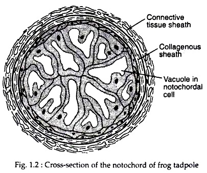
1. Notochord is present in any stage of the life-history. It is a slender rod that arises out of the dorsal wall of the embryonic gut in primitive chordates. Notochord lies dorsal to the coelom and parallel beneath the central nervous system. The notochord is composed of a core of cells and fluid encased in a tough sheath of fibrous tissue (Fig. 1.2). Notochord provides mechanical support and lateral flex to the body.
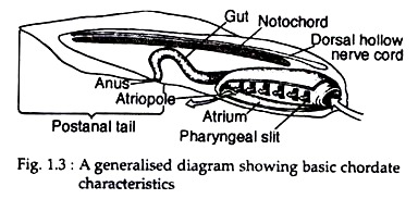
2. Pharyngeal gill slit (Fig. 1.3) is one of the fundamental features of the chordates. In the digestive tract, immediately posterior to the mouth, a spacious cavity is called pharynx. During some point in the life-cycle of all chordates, the walls of the pharynx are pierced or nearly pierced, by a lateral series of openings.
ADVERTISEMENTS:
These openings are called pharyngeal gill slits. In vertebrates, gills form adjacent to these pharyngeal slits. The slits are simple openings only, with no significant role in respiration. In many primitive chordates, these openings serve primarily in feeding.
3. Dorsal hollow tubular nerve cord (Fig. 1.3) is derived from the embryonic ectoderm by a process of invagination. Future nerve tube cells of the early chordate embryo gather dorsally into a thickened neural plate within the ectodermal surface. This plate invaginates to form nerve cord. It is to be noted that the major nerve cord in most invertebrates is ventral in position, (i.e. below the gut) and solid.
In chordates, the nerve cord lies above the gut and is hollow along its entire length. The hollow cavity is called neurocoel and it is fluid-filled. The advantage, if any, of a tubular rather than a solid nerve cord is not known, but this distinctive feature is found only among chordates.
4. Posterior elongation of the body extending beyond anus, is called the post anal tail. The tail is primarily an extension of the chordate locomotory apparatus, the segmental musculature and notochord.
5. Circulatory system is closed type. Heart is ventral in position and helps in circulation of blood with the aid of muscular contraction and relaxation. Generally haemoglobin is the respiratory pigment and present in blood.
6. Blocks of muscles called myomeres are sequentially arranged as external body wall. Internally different organs are arranged metamerically.
7. All the organs are developed from ectoderm, mesoderm and endoderm. Development of coelom is enterocoelic.
ADVERTISEMENTS:
8. Majority of higher chordates possess endoskeleton and exoskeleton. Endo- skeleton develops from mesoderm.
9. In the bi-laterally symmetrical body, anteroposterior axial organisation is clearly visible. Head is situated at the anterior end and tail in the posterior end.


