In this article we will discuss about the nervous system of rohu fish (Labeo rohita) with the help of suitable diagrams.
Brain:
The brain of Labeo, though in general not unlike that of elasmobranchs, is very different in detail and specialised. The cerebellum is very large. Its anterior part does not bulge outward. Instead it pushes forwards under the roof of mesencephalon to form valvula cerebelli. It is characteristic of teleost fishes. The optic lobes are also large with two large lobi inferiores on the ventral side.
The optic nerves do not form a chiasma, but simply cross one another or decussate. The diencephalon is much reduced and dorsally indicated only as the place of origin of the pineal body. The infundibulum between the lobi inferiores gives attachment to the pituitary body (hypophysis).
The small undivided prosencephalon comes immediately in front of the midbrain. Olfactory bulbs are situated in close apposition with the forebrain, without intervening olfactory tracts. Each contains a cavity (rhinocoele) communicating with the undivided ventricle of the forebrain.
ADVERTISEMENTS:
Three transverse bands of fibres connect the right and left halves of the forebrain, an anterior commissural joining the corpora striata, a posterior commissure situated just behind the origin of the pineal body, and an inferior commissure in front of the infundibulum.
The pineal body is rounded and placed at the end of a hollow stalk. No trace of any optic structure remains within. A shorter offshoot of the root of the diencephalon may perhaps represent a rudimentary pineal eye. Behind the pituitary body is a saccus vasculosus. It may be a pressure receptor.
Cranial Nerves:
There are ten pairs of cranial nerves and a pair of terminal nerves (cranial nerve, “O”). Their origin, distribution and nature are like that of Scoliodon.
ADVERTISEMENTS:
Spinal Nerves:
These are paired and each like that of Scoliodon arises as a dorsal sensory root and a ventral motor root.
Autonomic Nervous System:
In teleosts paired symmetrical chains of sympathetic ganglia begin at the terminal level and run backwards to the end of the tail. A ganglion is connected with each cranial dorsal root. These receive pre-ganglionic fibres from the ventral roots in the trunk region and pass forwards in the sympathetic chain. The sympathetic trunk is connected with the mixed spinal nerves by rami communicants.
Each ganglion receives a white ramus containing pre-ganglionic fibres from its spinal nerve and gives a grey ramus to the same nerve. The grey ramus carries postganglionic fibres, some of which are concerned with melanophore contraction. Thus, the autonomic nervous system is far more advanced than that of cartilaginous fishes. It is comparable to the basic plan in tetrapods.
Sense Organs:
ADVERTISEMENTS:
Olfactory receptors are paired olfactory pits, each of which opens to the external by means of two apertures but they do not communicate to the buccal cavity. Olfactory pits are lined by the olfactory cells. Their function is olfactory and has no role in respiration.
Taste buds (chemoreceptors) are found in many parts of the body. Taste buds are present over the lips, in the epithelial lining of the first three gill-slits and on the barbels.
Tactile receptors are abundant over the lips and barbels and are also found over the entire body.
Lateral line system is well developed in actinopterygians. Each sensory organ lies in a pit and communicates with surrounding water through a pore. These pits are linked by canals and innervated by the lateral line branch of the vagus. Lateral line system helps the fish to perceive low frequency vibrations in water and also apprises the fish of the approach of predator or prey.
Eye:
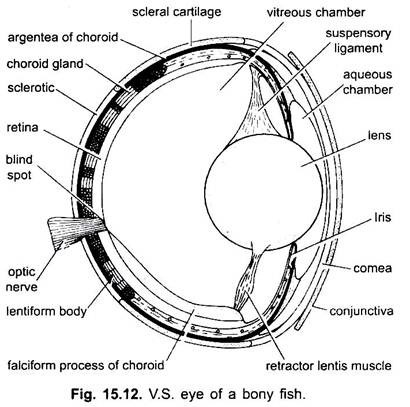
A pair of eyes over the head are the photoreceptors.
Each eye has three layers:
(i) Outer cartilaginous sclerotic layer,
(ii) A median vascular choroid layer and
ADVERTISEMENTS:
(iii) An innermost photosensitive retinal layer.
In between the sclerotic and choroid is a silvery layer or argentea which gives its colour. The cornea is flat with which the globular lens is almost in contact, so that the anterior chamber of the eye is extremely small.
There are no choroid processes. In the posterior part of the eye, between the choroid and the argentea, is a thickened ring-shaped structure, the choroid gland which surrounds the optic nerve. It is not glandular, but it is a complex network of blood vessels or rete mirabile. Close to the entrance of the optic nerve a vascular fold of the choroid, the falciform process, pierces the retina, and is continued to the back of the lens.
Here it ends in a muscular knob, the campanula Halleri or retractor lentis. The falciform process and campanula Halleri takes an important part in the process of accommodation by which the eye becomes adapted to forming and receiving images of objects at various distances. Accommodation is also effected by shifting the position of lens and not by changing the shape of lens as occurs in higher vertebrates. The pupil size appears to alter very little or not at all. Vision is monocular, each eye has different visual field.
Ear or Organ of Audio- Equilibrium:
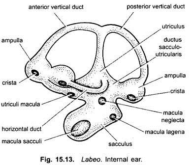
In fishes, the external and middle ears are absent, only internal ear or membranous labyrinth is found. The membranous labyrinth is formed of an upper utriculus and a lower sacculus. Three semicircular canals open into the utriculus. The sacculus is sac-like and its floor gives rise to a lagena.
The endolymph present in the membranous labyrinth contains otoliths or ear-stones, which are of three types- sagitta is relatively large and it almost fills the sacculus, asteriscus is a small granule lying in the lagena, and the lapillus is found in utriculus close to the ampullae of the anterior and horizontal canals.
Bony fishes hear with great discrimination, despite the absence of organ of Corti. The utriculus and the semicircular canals help the fish to maintain equilibrium. The sacculus and lagena perceive the sound waves. The body surface transmits the vibrations to the membranous labyrinth.

