In this article we will discuss about the external structures of pigeon.
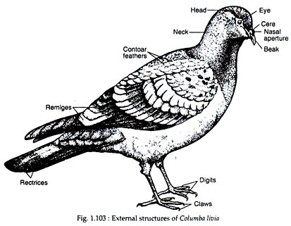
The size varies from 20-25 cm. The appearance varies in different varieties but all of them have a compact and more or less spindle-shaped body (Fig. 1.103). The body is distinctly divisible into three regions — head, neck and trunk.
(i) Head:
ADVERTISEMENTS:
The head is small, round and anteriorly pointed. It contains the following structures:
(a) Mouth:
It is a wide gap at the anterior end of head and bounded by upper and lower beaks. Both the beaks are covered by a sheath called horny sheath or rhamphothecae. The beaks are pointed, hard and suitable for its grain-eating habit.
ADVERTISEMENTS:
(b) Cere:
Near the base of the upper beak a pair of swollen, feather covered areas, called cere is present.
(c) External neres:
ADVERTISEMENTS:
Just anterior to the cere, a pair of external nares is present as minute slits.
(d) Eyes:
On each lateral side of the head, a prominent eye is placed. Each eye is provided with an upper, a lower and a movable transparent third eyelid or nictitating membrane, which runs across the eyeball.
(e) External ear:
These small paired apertures are present one on each lateral side of the head and slightly posterior to the eye. Each aperture remains covered by a special group of feathers called auricular feathers. Each aperture leads into a canal called the external auditory meatus.
(ii) Neck:
The neck is long and flexible. It holds the head over the trunk and its flexible nature is responsible for the universal movement of head.
(iii) Trunk:
The trunk is compact, stout and immovable. All the heavier organs are placed on the central part of the trunk and immediately beneath the wings.
ADVERTISEMENTS:
The trunk region contains the following structures:
(a) Forelimbs:
Paired forelimbs are connected with the anterior region of the trunk and are greatly modified as wings. At the time of rest, the wings lie on the lateral side of the trunk as a folded ‘Z’. Each wing has three divisions — upper arm, forearm and hand.
Folds of skin extend between upper arm and trunk and also between upper arm and forearm. The former fold is called post- patagium and the latter is known as pre-patagium. Both the patagia are greatly reduced. Each hand has three clawless insignificant digits.
(b) Hind limbs:
Paired hind limbs work as legs and are modified to carry the entire weight of the body during perching and walking on land. It is connected with the trunk near its posterolateral side. The thigh portion of the hind limb runs parallel to the body and during rest remains under the cover of the wings.
Each hind limb is provided with four toes of which the first one is called hallux which is directed backwards and the remaining three are pointed anteriorly. The upper part of the hind limb is feathered but the lower part is covered by scales.
All the toes are provided with sharp and pointed claws. At the time of flight, the hind limbs are kept withdrawn and are released like the wheels of an aero plane immediately before landing.
(c) Tail:
Posteriormost conical end of the trunk is known as uropygium. On the ventral side of the uropygium lies a transverse slit called vent or cloacal aperture, which serves as the outlet of cloaca. On the middorsal region of the uropygium there is a gland called uropygium or preen or oil gland.
This gland produces an oily secretion, which is drawn by the beaks and is sprayed all over the body to ‘dress’ the feathers. The uropygium has special series of feathers called rectrices. These feathers in the uropygium constitute the tail of pigeon. The tail is used as steering and brake during flight and works as balancing organ during walking or perching or flying.
(d) Skin:
The skin of pigeon is dry, thin and loose in texture. Skin glands are absent. The uropygial gland is the only integumentary gland. All over the skin numerous free nerve endings remain scattered. Near the base of the upper beak these nerve endings form specialised organ for touch in the cere.
In spite of its thin appearance, the skin has great ability to produce keratin, which is used largely in the formation of various integumentary derivatives.
(e) Skeletal Structures:
Pigeon possesses well-formed exoskeletal and endo-skeletal structures.
(i) Exoskeleton:
Following integumentary structures are seen in birds which perform different functions:
(a) Beaks:
These are horny exoskeletal derivatives which cover the upper and lower jaws. It is used during ingestion, preening of feathers and fighting.
(b) Claws:
Claws are pointed, sharp and horny exoskeletal organs which are present at the extremities of the toes. The claws are typically reptilian in construction and help during perching and walking.
(c) Scales:
Scales are reptile-like exoskeletal structures present on the exposed parts of the hind limbs. These scales originate from the epidermal part of the skin and remain arranged in tile-like fashion.
(d) Feathers:
These exoskeletal structures are the unique features of birds and are not seen in any other animal. These distinctive epidermal structures furnish a flexible, lightweight and resistant body covering of pigeon and are supposed to be modified reptilian scales. They cover most parts of the body and form the wings and tail.
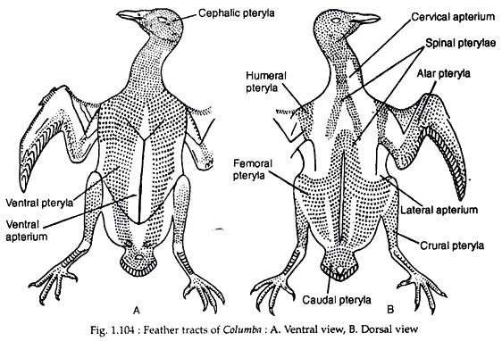
Each feather originates from the epidermal layer of the skin and are arranged in definite tracts called pterylae, which are separated from the featherless tracts called apteria (Fig. 1.104). The arrangement of feathers on the body of pigeon is called pterylosis. The assemblage of feathers at one time is called the plumage.
Feathers may be white (due to the presence of air spaces) or coloured (due to the presence of pigments). The colour of the feathers is due to chemical pigments and the physical arrangement of the elements composing the feathers. Feathers are periodically replaced by a process called moulting, which occurs sporadically throughout the body.
Kinds of Feathers:
The feathers of pigeon may be classified into two groups:
(A) Flight Feathers or Quills:
These feathers are associated with flight and may again be subdivided into wing feathers or remiges and tail feathers or rectrices. The remiges are present in the wings and each wing has 23 feathers, which are again sub-divided into primaries, secondaries and tertiaries.
The remiges attached to the distal part (carpometacarpus) of the wing are primaries, those on the middle (radius and ulna) portion are secondaries and those attached to the proximal part (humerus) of the wing are called tertiaries. The rectrices are twelve in number and are arranged semi-circularly in the tail region.
(B) Covering Feathers:
These are responsible for the general covering of the body and are of three types: Contour feathers, Filo-plumes or hair feathers and Down feathers or plumules.
The contour feathers form the majority of the covering feathers and filo-plumes remain in between the contour feathers.
The down feathers are also present in- between the contour feathers. In the body of the nestling it is called powdered down feather.
Structure of a typical feather:
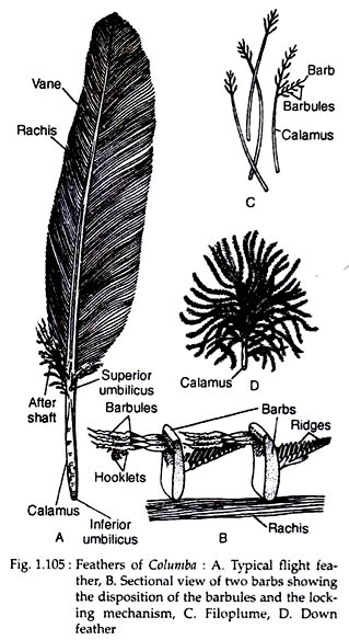
The feathers differ in their size, shape and function, but are built up in the same general plan. A typical feather has the following two parts — shaft and vane or vexillum (Fig. 1.105A).
The shaft portion is divisible into two parts, calamus (Gk. kalatnos, quill) and rachis (Gk. rakhis, spine).
Calamus or the Quill:
The calamus is the hollow cylindrical stalk by which the feather remains attached with the skin. At the proximal end, there is an aperture called inferior umbilicus into which fits the mesodermal papilla of the skin. At the distal end, there is another aperture called superior umbilicus.
Rachis:
The rachis is solid pith-like in appearance and is continuous with the calamus. A small tuft of soft feathers, called the after-shaft, develops near the superior umbilicus. The vane or vexillium represents the broad part of the feather.
It is formed by delicate closely set structures, called barbules or rami, which extend from each side of the rachis. The distal edge of each barb has two sets of barbules—proximal and distal. The proximal barbule from one barb crosses the distal barbule of the next.
The tips of the distal barbules are provided with hooklets and the tips of proximal barbules have flanges. The hooklets and flanges interlock to render a compact surface of the vane which resists the passage of air. The after shafts are few hairy processes present just proximal to the superior umbilicus.
In the flight feather, the shaft is long and vanes are broad. Among the covering feathers, the contour feathers have short shaft but extended vane portion. Each filo plume has a long shaft carrying a few degenerated barbs and barbules at the tip and such barbs are without interlocking apparatus.
Similarly, the down feathers are also without interlocking apparatus in the barbs. The down feathers consist of a short quill or stem. The barbs are arranged in a circle at the top of the stem. The barbs have minute barbules. Each feather is provided with a muscle at its base, which controls the position of feather.
Uses of feathers:
(i) The nerve fibres present at the base help the feather to act as a sensory structure.
(ii) The feathers produce a compact surface to resist air-current.
(iii) It retains heat and thus helps the bird from the temperature hazards.
(iv) It is light and, therefore, helps in flight.
(v) In some birds, a few feathers act as secondary sexual characters.
Development of feather:
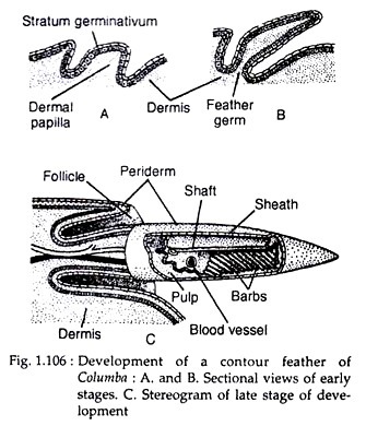
The germ of a developing feather begins as a dermal papilla of the skin composed of epidermis and dermis (Fig. 1.106A, B). At the initial stages, the dermal cells push the overlying epidermis up ahead of it. The base of the outgrowth gradually sinks into the skin.
The outgrowth includes dermis and epidermis. The epidermis becomes hard and cornified and is pushed out from the papilla region. The dermal plug gradually withdraws leaving a hollow quill inserted in the skin.The embryonic feather is, thus, a tube of cornified epidermis set in a pit of dermis.
One side of tubular embryonic feather becomes considerably thickened to transform into the shaft (Fig. 1.106C). From the shaft slanting barbs are developed on either side. The wall of the tube opposite the shaft becomes very thin. Rupture of this thin portion causes the spreading out of the rolled-up feather to assume its usual form.
(b) Endoskeleton:
Pigeon possesses well-developed endoskeleton. The entire endo-skeletal frame may be grouped into axial and appendicular skeletons. Both these groups have undergone considerable modifications, specially fusion of different parts, to meet the requirements of the birds peculiar way of life.
(i) Axial skeleton:
The axial skeleton includes the skull, vertebral column and sternum.
(a) Skull:
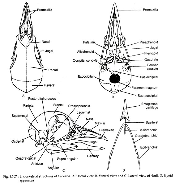
The skull of pigeon is light and fragile. It is more or less round in shape. The skull bones are paper-like thin. The sutures of the skull bones are obliterated. The foramen magnum is directed ventrally and the skull articulates with the vertebral column by a single round occipital condyle on the anterior border of the foramen magnum.
The cranial part of the skull is formed dorsally by a pair of parietals and a pair of frontals (Fig. 1.107A). The ventral side of the cranium is formed posteriorly by basioccipital and anteriorly by basisphenoid. The basisphenoid is drawn anteriorly as a narrow rosturm. Ventrally, the basisphenoid gives rise to two membranous basitemporals which fuse to form the posterior part of the presphenoid.
The anterior part of the presphenoid is represented by the rostrum. The supraoccipital and the exoccipital form the posterior and lateral sides of the cranium (Fig. 1.107B). On each side of the cranium lies the tympanic cavity. It is formed by the squamosal, prootic and epiotic bones.
Two orbits are separated by inter-orbital septum, which is formed by the union of presphenoid and mesethmoid bones. The nasals are forked in appearance and form the inner and outer margin of the nasal cavity. A large lacrymal bone is present on each side between the orbit and the facial part of the skull.
The facial part of the skull is united with the cranial part in the following way: maxilla unites with the jugal and quadrato-jugal and ultimately with the quadrate. Quadrate is a three-rayed bone which articulates with the tympanic cavity by two processes and sends an orbital process. It bears condyle for the articulation of the mandible. The palatine articulates with the pterygoids and rostrum posteriorly.
The pterygoids articulate with the quadrate and also with the basipterygoid process of the rostrum. The vomer is absent in pigeon. The maxillopalatines remain separate and do not unite medially. This particular type of the skull is called schizognathous type of skull. The mandible consists of dentary supra-angular, angular, splenial and articular (Fig. 1.107C).
Hyoid apparatus:
The hyoid apparatus (Fig. 1.107D) consists of an arrow-shaped basihyal from which two short cornua originate. It is continued as slender basi-branchial from which originates a pair of posterior cornua. Each cornu contains ceratobranchial and epibranchial segments.
Dorsally, the hyoid apparatus is represented by a slender bone called columella which remains united with the stapes of the middle ear by one end and the other end has three cartilages, extra-stapedial, infrastapedial and supra-stapedial, to fit into the tympanic membrane.
Vertebral Column:
The vertebral column is an elongated structure. It shows flexibility at the anterior part, compactness at the trunk region and shortening near the tail region. Entire column is divisible into cervical, thoracic, synsacral, caudal and pygostyle regions. Each region includes several vertebrae.
(a) Cervical vertebrae:
Altogether 14 cervical vertebrae support the neck region of pigeon and are constructed in such a way that, while supporting the head, they permit free movement of the neck.
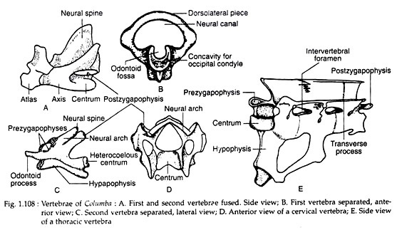
A typical cervical vertebra (Fig. 1.108B, C, D) has the following features:
(i) Centrum is heterocoelous (anteriorly concave from side to side but convex from above downwards and posteriorly concave from above downwards but convex from side to side). The heterocoelous centra of the neck vertebrae give greater mobility of neck and allow the head to move at an angle of 180°.
(ii) Articulating surface of the centrum contain synovial capsules, cartilaginous plates, meniscus and suspensory ligament.
(iii) Double-headed vestigial ribs are directed posteriorly.
(iv) The transverse processes are perforated to allow the passage of vertebral artery; this passage is called the vertebrarterial or transverse foramen. The first two cervical vertebrae differ from a typical cervical vertebra in structure.
In both, the different parts are much reduced. The first vertebra, which is also known as atlas, is a small ring-like structure which remains fused with the second vertebra called axis. (Fig. 1.108A, B).
(b) Thoracic vertebrae:
Four to five in number, of which the last one is free and the rest are united. The last thoracic vertebra is fused with the synsacrum. In each thoracic vertebra, the centrum is ventrally compressed into a plate called hypophysis (Fig. 1.108E).
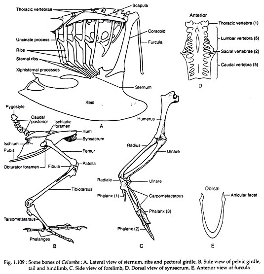
A pair of ribs remains attached with the vertebra by two heads—one head is united with the centrum and the other head remains united with the transverse process. This vertebral portion of the rib is attached with a bony counterpart from the sternum. Bony interconnection — called uncinate processes — interlocks with posterior ribs to make the thoracic box rigid (Fig. 1.109A).
(c) Synsacrum:
It is a compact more or less triangular skeletal mass formed by the fusion of vertebrae (Fig. 1.109D). Together with pelvic girdle, it renders rigidity to the body.
Following vertebrae constitute the synsacrum:
(i) Posterior thoracic—only one with ribs not connected with the sternum.
(ii) Lumbar—Five to six in number. Ribs are absent. Transverse processes are placed high on the neural arch and their uniting ligament is ossified to form a dorsal continuous bone.
(iii) Sacral—Two in number. Transverse processes of each sacral originate near the neural arch and the centrum is provided with a ventral outgrowth which lies pressed against the pelvic girdle.
(iv) Anterior caudal—Five in number, and all are fused with the other parts of the sacrum.
(d) Posterior caudals:
Six in number. Each one is free and distinct (Fig. 1.109B). Each vertebra is with a short and prominent centrum.
(e) Pygostyle or Ploughshare bone:
It is formed by the fusion of last four vertebrae (Fig. 1.109B). The pygostyle is a compressed skeletal structure and is turned upwards.
Sternum:
The sternum is extremely modified due to the flying habit. It is modified ventrally into a boat-shaped bony skeletal structure called keel or carina (Fig. 1.109A). The anterior part is provided with a pair of grooves for the attachment of the coracoid bones of the pectoral girdle. The posterior part possesses two pairs of notches.
Appendicular skeleton:
It consists of skeletal elements which constitute limbs and girdles. Among the limbs, the forelimbs are modified as wings and the hind limbs are modified as legs. The bony components of the forelimbs become greatly altered due to their transformation into wings. The pectoral girdle is built up for the attachment of wings and the pelvic girdle holds the hind limbs to support the body during standing.
Pectoral girdle and wings:
The appearance of the pectoral girdle is altogether different from that of other vertebrates.
The pectoral girdle of each side contains following parts:
(i) Coracoid:
It is a massive, rod-like strongly built bone. Each coracoid is placed on the dorsoventral axis and is attached to the broad end of the keel. Anterior end contains a process called acrocoracoid process.
(ii) Scapula:
It is a sabre-shaped bone which extends anteroposteriorly over the ribs (Fig. 1.109A). At its anterior end, it is connected with the coracoid by a ligament. The coracoscapular angle is less than 90°. This is a characteristic feature of the flying birds.
(iii) Furcula or wish bone or Merry thought bone:
It represents the clavicles and inter-clavicles of pectoral girdle of other vertebrates. It forms a ‘V’-shaped structure and hangs along the dorsoventral axis in front of the sternum (Fig. 1.109E). A small aperture (foramen triosseum) is left at the junction of the coracoid, scapula and furcula. Through this aperture, the tendon from the wing muscles passes for its attachment with the humerus.
(iv) Glenoid cavity:
It is the place where the head of the forelimb fits in. It is formed by both coracoid and scapula, the latter forms acromion process within it.
Each wing contains following skeletal pieces (Fig. 1.109C):
(i) Humerus:
It is a strongly built bone with broad head. The head bears a number of prominent ridges and a pneumatic foramen.
(ii) Radius and Ulna:
Both the bones are separated in the middle but remain closely apposed at the two ends. Radius is a slender, straight skeletal piece, while ulna is curved and slightly bent.
(iii) Proximal carpals:
Two large skeletal pieces, one called radiale and the other called ulnare — remain free.
(iv) Carpometacarpus:
This small but compact bone includes two skeletal pieces, which are fused at both the ends but remain separated in the middle. One piece is stout and straight, while the other piece is slender and curved. It is formed by the union of two distal carpals and three metacarpals.
(v) Phalanges:
Following phalanges are present in each limb, all of which are devoid of claws.
They are:
(i) One pointed phalanx is attached with the first metacarpal.
(ii) Two phalanges remain attached with the second metacarpal, one of them is flange-like and the other one is pointed.
(iii) One pointed phalanx is attached with the third metacarpal (Fig. 1.109C).
Pelvic girdle and hind limb:
The pelvic girdle is compact and ankylosed with the synsacrum.
It contains following skeletal parts on each side:
(i) Ilium:
It is a broad and large bone, fused with the synsacrum on its inner side. It is concave anteriorly and convex posteriorly.
(ii) Ischium:
It is a broad bone, which is fused with the ilium at its posterior end. Anteriorly, an ischiatic foramen separates the ilium from ischium. No ischiatic symphysis is present.
(iii) Pubis:
It is a narrow, curved and elongated skeletal piece, which is placed parallel to the ischium and separated from it by obturator foramen. No pubic symphysis occurs in pigeon.
(iv) Acetabulum:
It is the cavity where the head of the femur fits in. Its dorsal side is formed by the ilium and the remaining part is formed by equal share from ischium and pubis. The edge of the cavity is drawn into a process called anti-trochanter, which works against the trochanter of the femur of hind limb (Fig. 1.109B).
Each hind limb is composed of following skeletal pieces:
(i) Femur:
It is a short and narrow skeletal piece. Its proximal end contains pointed trochanter and rounded head. The hind limb is placed parallel to the sagittal plane of the body. The distal end is drawn into a pulley-like condyle, containing a patella as knee-joint.
(ii) Tibiotarsus:
It is a long bone formed of tibia with the fusion of two distal tarsals.
(iii) Fibula:
It is a small rudimentary bone situated on the lateral surface of the tibiotarsus.
(iv) Tarsometatarsus:
This bony segment is formed of some of the tarsals and the metatarsals of the second, third and fourth digits. A small vestige of the first metatarsus is located on the posterior side of the distal end of the tarsometatarsus. The ankle joint of bird — being mesotarsal — resembles that of reptiles.
(v) Phalanges:
Pigeon has four digits; the fifth one is lacking. Phalanges are present in the following number at different toes and the distal phalanges of all the toes are provided with sharp and pointed claws. The first toe or hallux has two phalanges and the second, third and fourth toes have three, four and five phalanges, respectively.
