In this article we will discuss about the respiratory system of pigeon.
The aerial mode of life requires extra energy. In order to get energy, pigeon eats large quantity of food and to break down the assimilated food at a faster rate, the respiratory system is extensively modified. The lungs are proportionately smaller in size, but the functional efficiency is greatly increased by the development of air-sacs.
The respiratory system of pigeon has two unique features:
(1) Presence of non-elastic, compact lungs
ADVERTISEMENTS:
(2) Possession of several air-sacs.
Following structures are present in the respiratory system of pigeon:
1. External nares:
ADVERTISEMENTS:
These are paired openings, present near the base of the upper beak and within the cere.
2. Internal nares:
Single opening which opens at the roof of pharyngeal region and both the external nares communicate through this common aperture.
3. Glottis:
ADVERTISEMENTS:
This is a slit-like aperture, which is present on the floor of the mouth cavity and near the base of the tongue. It leads into the next part called trachea.
4. Trachea:
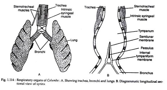
This elongated tube begins from glottis and runs along the neck region along the ventral side of the oesophagus. The trachea is composed of complete bony tracheal rings (Fig. 1.114). Near its commencement, the trachea is enlarged into a chamber called the larynx.
This chamber is supported by a cricoid cartilage (which is composed of four pieces) and one pair of arytenoid cartilages. In birds, the larynx does not function as voice box.
5. Syrinx:
Near the junction of neck and trunk, the trachea is swollen into a chamber called syrinx (Fig. 1.114B). It is formed by the dilatation of the last three or four tracheal rings and first bony ring of each bronchus.
The mucous membrane of the syrinx constitutes a pad-like thickening and is provided with several muscles and membranes. The syrinx is actually the voice box. The syrinx is the characteristic organ of pigeon and many other flying birds.
It has a complicated structural construction. A bar of cartilage called pessulus is present at the junction of two bronchi. It extends dorsoventrally inside the tympanum and holds a small fold of mucous membrane called semilunar membrane or membrana semilunaris (Fig. 1.114B).
ADVERTISEMENTS:
The inner membranous lining of the bronchi produces inconspicuous internal tympani-form membranes. Sound is produced by the vibration of the membrana semilunaris while the pitch of the sound is controlled by the action of the syringeal musculature.
The syringeal musculature includes a pair of intrinsic syringeal muscles (which arise from the lateral sides of the trachea and are attached with the syrinx) and a pair of sternotracheal muscles (originate from the sternum and are inserted into the trachea). The position of syrinx can be changed by the action of these muscles.
6. Bronchus:
Within the trunk, the trachea bifurcates into right and left bronchi. The first ring of each bronchus is complete and bony, while the rest are incomplete mesially. The left and right bronchi are called primary bronchi or mesobronchi. Each mesobronchus, in the beginning, is composed of rings of cartilages, but inside the lung such rings are absent.
Each primary bronchus enters the lung through a small space called vestibulum and extends up to the posterior extremity of lung. Within the lung, the mesobronchus sends a pair of branches called secondary bronchi and each secondary bronchus breaks up into a network of tertiary or para-bronchi and sends branches to the air-sacs.
Each tertiary bronchus again subdivides into numerous finer network of tubules (air capillaries), which remain in close contact with the blood capillaries. While running posteriorly, the diameter of mesobronchus gradually decreases and it finally opens into the abdominal air-sac by an ostium.
7. Lungs:
The lungs are small in size in comparison with that of the body. These are paired pink-coloured organs. The lungs are spongy organs with little elasticity. The dorsal surface of the lungs is fitted closely with the interspaces of ribs and lacks the peritoneal covering, i.e., pleura is absent on the dorsal side.
The ventral wall has a compact fibrous tissue sheet called pleura or pulmonary Apo neurosis. The wall of the pleura has special fan shaped costopulmonary muscles which originate from the joints of vertebral and sternal ribs. The alveolar lining is formed by the ramification of tertiary tubules with the distribution of blood vessels.
8. Air-sacs:
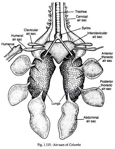
The air-sacs are bladder-like structures. These are formed by the dilation of the mucous membrane of the bronchus. The air-sacs are thin-walled membranous sacs and are devoid of blood vessels. Following air-sacs are present in the body of pigeon and all of them remain in communication with the pneumatic cavities of bones. There are nine major and four accessory air- sacs in pigeon (Fig. 1.115).
Major air-sacs:
The major air-sacs (Fig. 1.115) originate directly from the lungs. Of the nine air-sacs, four are paired and one is unpaired:
(a) Paired air-sacs:
(i) Posterior or abdominal air-sacs.
(ii) Posterior thoracic air-sacs.
(iii) Anterior thoracic air-sacs.
(iv) Cervical air-sacs.
(b) Unpaired air-sac:
(i) Inter-clavicular or median air-sac.
Accessory air-sacs:
These air-sacs originate as paired diverticula from the inter-clavicular air-sac.
These paired sacs are:
(i) Clavicular air-sacs, and
(ii) Humeral air-sacs.
Inspiratory air sacs:
1. Abdominal air-sacs:
These are also called the posterior air-sacs and lie among the coils of intestine. These are the posterior- most and largest air-sacs in birds. The right air-sac is larger than its left counterpart. These air-sacs send diverticula into the pelvic girdle, synsacrum, hind limbs and between thigh muscles.
2. Posterior thoracic air-sacs:
These paired air-sacs are placed on the posterior side of the thoracic cavity. The left sac is slightly larger than the right. Both the sacs are closely apposed with the lateral wall of the body cavity. Each bronchus, near its entrance into the lung, gives three short branches: One enters into the anterior thoracic air-sac, the second is connected with the cervical air-sac and the third enters the inter-clavicular air-sac.
3. Anterior thoracic air-sacs:
These paired air-sacs are located one on each side of the thoracic cavity towards the anterior part between the lungs and the ribs.
Expiratory air sacs:
4. Cervical air-sacs:
These paired air- sacs are placed near the base of the neck and lie in front of the lungs. Each sac sends diverticula into the cervical vertebrae and the skull.
5. Inter-clavicular air-sac:
This is an unpaired and median air-sac of large size. It has two ducts, one opening into each lung. Although this sac is unpaired in adult, it is formed by the fusion of two sacs which are evident by the presence of two ducts.
Each side of this air-sac gives off two extensions—
(i) Clavicular air-sac and
(ii) Humeral air-sac.
These sacs are communicated with the cavities of the bones.
A layer of fibrous tissue called oblique septum encloses the ventral walls of both the thoracic air-sacs. This septum extends up to the pericardium and unites with the similar septum of the other side. Such union along the middle line separates the body cavity into two chambers.
One chamber houses the lungs, thoracic and inter-clavicular sacs and the other chamber contains the heart, liver, stomach, intestine and abdominal air-sacs.
Role of air-sacs:
The air-sacs play an important role in the life of flying birds. The air-sacs are not provided with capillary network, so they are not directly respiratory in function. Besides, these sacs are essential components for aerial life.
The different uses of air-sacs are:
Function as bellows:
As the lungs are anchored firmly to the dorsal wall of the thoracic cavity, the elasticity of the lungs is greatly hampered. To carry on an efficient circulation of air through lungs during respiration, some mechanical aid becomes necessary, specially during flight. The air-sacs give mechanical aid by acting as the bellows.
Act as balloons:
When the air-sacs are inflated due to intake of warm air, the specific gravity of the body is lowered to a considerable extent. As the warm air is lighter than ordinary air, the retention of such air inside the air-sacs makes the body considerably lighter. This also lessens muscular efforts to sustain the body heavier than air.
Function as ballast:
The air-sacs are so nicely arranged on the two sides of the body that proper centre of gravity is established for balanced flight. If the equilibrium is lost by chance during flight, restoration of the equilibrium is easily maintained by shifting of the contained air from one side of the body to the other.
Lessen mechanical friction:
The air-sacs send branches which are inserted between the muscles (specially the flight muscles) like pads. Such a placement of air-sacs reduces mechanical friction to a large extent and increases the flexibility of the wings during flight.
Regulate and maintain body temperature:
The skin of bird lacks integumentary glands. So the skin has no utility in the regulation and maintenance of body temperature. Retention of warm moist air inside the air- sacs helps to regulate and maintain the body temperature.
Serve as the containers of reserve air:
During rest, respiration involves the alternate depression and elevation of the breastbone caused by the activity of the intercostal muscles. But during flight the breastbone as well as the thoracic basket is kept in a rigid state and the intercostal muscles remain in tension.
The respiratory process is slightly hampered for the time. Therefore, some internal source of reservoir of air becomes indispensable. The air-sacs sub serve this function and ventilate the lungs during flight.
Act as resonator:
The pitch of the sound is controlled to some extent by the forceful expulsion of the air from the air-sacs which act as resonator.
Regulate the moisture content of air:
Water is evaporated from the walls of the air- sacs in birds. So the air-sacs regulate the water content of the body.
Mechanism of respiration:
The unique feature of avian respiration is the double supply of oxygenated air to the surface of lungs for improved aeration. For this reason, respiration in birds is called double respiration. Two cycles of inspiration and expiration are required for a single volume of gas to move through the respiratory system. During the first inspiration, the sternum is lowered, and the lungs and the air-sacs are expanded.
The fresh air rushes through trachea and bronchi into the posterior air sacs. During the first expiration, the sternum is raised and the air from the posterior sacs goes to the Para bronchi and air capillaries. At second inspiration, air from the Para bronchi goes to the anterior air sacs and, on the second expiration, the air is expelled outside.
In other air-breathing animals, in between inspiration and expiration, some amount of residual air always remains within the cavity of lungs. But in birds, the residual air remains inside the air sacs and in the smaller branches of the bronchi. As the aeration of blood is complete in pigeon, it thus increases the respiratory efficiency to yield extra energy.
The respiratory movements are caused by two sets of muscles; one set operates during flight and the other set works at the time of rest. During flight, both inspiration and expiration are caused by the movement of pectoral muscles. At the time of rest, inspiration is caused mostly by the activities of intercostal muscles and expiration by the movement of abdominal muscles.

