
In this article we will discuss about the external structures of calotes. This will also help you to draw the structure and diagram of calotes.
The body of Calotes is divisible into three regions: head, trunk and tail (Fig. 1.85). A small narrow neck joins the head with the trunk. The head is more or less triangular in appearance. The snout is short, pointed and bears a pair of apertures called external nares. The eyes are provided with movable nictitating membrane. Mouth is a transverse aperture and is terminal in position.
Lips are absent. The tympanum is situated behind the eyes. Trunk is elongated, sub- cylindrical and bears two pairs of appendages, the forelimbs and hind-limbs. Each forelimb is divisible into brachium, anti- brachium and manus. Likewise, the hind- limb is divisible into femur, crus and pes. The limbs end in digits and the digits are provided with sharply pointed claws.
The cloacal aperture or vent is a transverse opening and is situated in the posteroventral part of the trunk and at the base of the tail. A large cloacal plate is situated in front of the cloacal aperture. The tail is long, slender and tapering. It measures twice as long as the head and body. Males measure 120-140 mm from snout to vent with a tail of 300-500 mm. The body length of females is less by 15-20 mm.

The colour of the body is brown or greyish on the dorsal surface and dirty whitish ventrally. The dorsal surface may be uniform in colour or may have dark-brown transverse spots or bars. Dark streaks radiate from the eye region. Young females often show two light yellow dorsolateral stripes. Fully grown males have greenish tinge. The tail exhibits alternate dark and light annuli.
Skin:
The skin of Calotes is highly modified for living on land. To safeguard the animal against the frictional contact with the dry ground as well as to prevent desiccation in dry air, the epidermis becomes greatly elaborated than the dermis. The only skin glands recorded in Calotes are the femoral glands in males.
These are present along the posteroventral margin of the thigh. These glands become functional during the reproductive period. The absence of skin glands and acquisition of scales over the body are evolutionary achievements for living on dry land.
ADVERTISEMENTS:
The skin of Calotes develops first as a simple cuboidal ectoderm. This ectoderm then differentiates into an outer periderm and an inner basal germinative layer. The epidermis becomes stratified due to addition of outer layers and the scales appear subsequently.
The epidermis is a compound layer which is composed of an inner layer of columnar cells called stratum germinativum (Malpigian layer) which is arranged on a basement membrane (Fig. 1.86A).

The outermost layer of epidermis is represented by a dead startum corneum. Between the stratum corneum and the stratum germinativum lies the transitional layer. The stratum corneum becomes thickened over the scales, while between the scales this layer becomes thinner.
ADVERTISEMENTS:
Skeletal Structures:
Calotes has definitive exoskeletal structures outside the body and well-developed framework of bony endoskeleton.
Exoskeleton:
Being a terrestrial animal, Calotes possesses well-developed exoskeletal structures. The body is covered by a non-conducting coat of keratin scales.
Scales:
The skin of Calotes is covered by scales. The scales form a continuous cover over the entire body of Calotes but become thinner in the grooves between the scales. Sensory structures in the form of sensory bristle (Fig. 1.87) are present on some large as well as small scales.
These sensory structures are called prototrichs (plural of protothrix). The prototrichs of reptiles have been claimed by many to be precursors of the mammalian hair. It is quite reasonable that the small scales margining the larger ones are the forerunners of feathers and hair.

The scales are epidermal in origin and are arranged in an imbricate fashion. The scales on the ventral surface of the body are smaller in size than those on the dorsal side. The scales develop from the stratum germinativum (or Malpighian layer) of the epidermis. These are plate-like structures, each is supported by a bony plate or ossicle or osteoderm (Fig. 1.86A).
ADVERTISEMENTS:
The osteoderms are tied into the dermis by Sharpeys fibers. On the dorsal side, the postaxial scales are larger than the pre axial scales. The scales on the head region are large and do not overlap but touch each other by their edges. These larger scales are called head shields.
Few scales situated on the mid-dorsal line and on the border line of neck have become modified into large and pointed structures. These are capable of movement and are usually called frills. Similar structures are also present on the posterolateral border of the head. The scale cover is periodically shed by a process called ecdysis.
Claws:
The digital tips of Calotes are provided with sharp claws. Each claw is made up of a dorsal and a ventral scale-like horny plates. These plates are so placed that they converge at the end of the digit to make the tip pointed.
The dorsal plate is called unguis. It is convex and rounded at tip and lateral sides. The ventral plate is called sub unguis. The sub unguis is flattened (see Fig. 1.86B). The plates are derived from the Malpighian layer of the epidermis. The unguis is more developed than the sub unguis.
Endoskeleton:
The hard skeletal parts present inside the body (endoskeleton) are grouped into axial and appendicular divisions. The axial skeleton includes the skull and vertebral column while appendicular skeleton includes the girdles and limb bones.
Skull:
The skull of Calotes is well-ossified but cartilaginous elements persist in the region of nares and inter-orbital septum. The brain box or carnium is comparatively small and is covered by investing bones.
The roof of the skull is made up of a pair of parietals which are fused and bear a median parietal foramen, a pair of frontals and two nasal bones (Fig. 1.88A). The premaxillae are situated anterior to and between the nasals while the sickle-shaped squamosals are attached to the posterolateral sides of the parietals by sutures.
On the roof of the orbit there are many small bones, of which the anterior and posterior ones are known as prefrontal and postfrontal, respectively. The floor of the skull is composed of a flat and large basal bone formed by the fusion of occipital and sphenoidal elements. The basal bone continues forward as a slender bar and is called Para sphenoid (Fig. 1.88B).
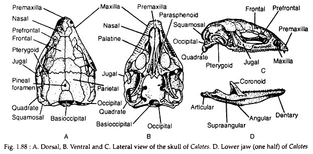
From the basal bone arise a pair of stout basipterygoid processes. These processes articulate with the pterygoid. The pterygoid forms a bridge between the quadrate (posteriorly) and palatine (anteriorly).
From the junction of pterygoid and palatine arises a stout os transversum or trans-palatine process which extends to join the maxilla. The vomer is small, anterior to palatine and articulates with premaxilla and maxilla. The premaxillae and maxillae are furnished with small teeth.
The posterior part of the skull is made up of large exoccipitals which are continuous with the horizontal prootic processes. The foramen magnum is an aperture situated between the occipitals through which medulla oblonata emerges as spinal cord. Single occipital condyle is present on the basi occipital.
The lateral wall of the cranium is mainly formed by a paired prootics. The epipterygoid (or columella) is extended from the prootic to the pterygoid. The jugal bones border the lateral and posterior limits of the orbit (Fig. 1.88C). The quadratojugal is not ossified.
The lower jaw is composed of two similar halves or rami. Each ramus, in addition to the slender persistent Meckels cartilage, consists of six pieces of investing bones which fuse with each other. The names of the six bones are: articular, angular, supra-angular, coronoid, splenial and dentary.
Of the six bones, the dentary has mandibular teeth (Fig. 1.88D). The two rami are sutured to one another in front. The lower jaw is articulated to the cranium through the quadrate which is movable and such attachment of lower jaw is streptostylic in nature. The lower jaw bears teeth.
Vertebral column:
The vertebral column consists of many vertebrae.
The vertebral column is divisible into the following regions:
(a) Cervical region—composed of 8-10 vertebrae,
(b) Thoracolumbar region—made up of 22 vertebrae,
(c) Sacral region—containing only two vertebrae and
(d) Caudal region— consisting of about 20 vertebrae.
All vertebrae, excepting the first and the last, are strongly procoelous.
(a) Cervical vertebrae:
The first cervical vertebra is known as atlas (Fig. 1.89A). It is ring-like having a single articular facet.

Centrum is absent. The neural arch is incomplete dorsally and transverse processes are ill-developed. The second cervical vertebra is called axis (Fig. 1.89B). It bears a small peg like process called odontoid process at its anterior face.
The odontoid process actually represents the centrum of the atlas. The mid- ventral line of the axis is provided with a ridge called hypapophysis. The rest of the cervical vertebrate are typical but bear cervical ribs. The atlas and axis do not bear ribs.
(b) Thoracic vertebrae:
The thoracic vertebrae are typical in nature (Fig. 1.89C, D). The hypapophysis is ill-developed or absent. All the vertebrae bear thoracic ribs. Only five anterior thoracic ribs reach the sternum.
(c) Sacral vertebrae:
The two large sacral vertebrae bear large and expanded ribs.
(d) Caudal vertebrae:
The ventral surface of the caudal vertebrae is provided with Y-shaped chevron bones.
Sternum:
The sternum (or breast bone) is a rhomboidal plate-like structure situated in ventral position of the thoracic box. The sternum is supported by a characteristic T- shaped interclavicle and it bears an aperture called sternal aperture at its middle (Fig. 1.90). The sternum bears ribs. The plate-like sternum protects the internal organs.
Hyoid apparatus:
The floor of the buccal cavity is supported by a cartilaginous structure called the hyoid apparatus (Fig. 1.89E). It also supports the tongue. The hyoid apparatus consists of basihyal, anterior cornu, middle cornu and posterior cornu. The basihyal constitutes the body proper of the hyoid apparatus.
It is a median cartilaginous rod and directed anteroposteriorly to form the os entoglossus. The anterior cornua are elongated cartilaginous rods and arise anterolaterally from the basihyal. These rods curve round the oesophagus and terminate near the ventral side of the auditory capsule. The anterior cornua represent the hyoidean arch.
The middle cornua are ossified at their proximal ends and represent the vestiges of the first branchial arches. The posterior cornua arise from the posterolateral edge of the basihyal. They extend backwards and outwards. Each cartilaginous rod is composed of two segments.
Appendicular skeleton:
The skeleton of the paired limbs and girdles constitutes the appendicular skeleton.
Pectoral girdle:
The pectoral girdle is hoof-shaped and composed of two symmetrical halves. It is situated in the thoracic region of the body, being attached to the vertebral column by means of muscles and ligaments. Each half of the girdle is made up of a partially cartilaginous scapula. The scapula is dorsolaterally extended (Fig. 1.90).

The suprascapula is a bony plate-like structure and is attached to the scapula. It is ventrolateral in position. The coracoid is also plate-like and is made up of cartilaginous epicoracoid and precoracoid. The glenoid cavity is formed by the scapula and coracoid. The clavicle is a narrow-curved bone and forms a bridge between inter-clavicular junction and scapula.
The two halves of the girdle are attached to a rhomboidal and plate-like sternum which is ventral in position. The sternum is supported by a T-shaped episternum (interclavicle). The sternum bears ribs and a centrally placed sternal foramen through which the sternal artery passes. The T-shaped interclavicle, sternal ribs and sterna framen are diagnostic features of the reptilian pectoral girdle.
Pelvic girdle:
The pelvic girdle is also made up of two equal halves. Each half is called os innominatum which is composed of pubis, ischium and ilium (Fig. 1.91). These three bones are attached in a tri-radiate fashion. The two halves of the girdle are attached with each other through the intermediation of pubis and ischium.

Hence the joining is called the ischiopubic symphysis. The pubis bears an epipubic cartilage at its tip and a pubic prominence as a projection at the other tip. The articulation facet, acetabulum is situated close to the pubic prominence. Between the two ischia there lies the hypoischiatic cartilage. The obturator foramen is incomplete.
Forelimb:
The forelimb consists of some long bones arranged in order from proximal to distal end (Fig. 1.92). Humerus. It is a slender and slightly curved bone which bears swellings (or condyles) at the proximal end. This swelling is called head of humerus. The distal end bears a pulley-like trochlea. Spherical epiphysis is associated with both ends. The deltoid ridge is absent.

The anti brachium is supported by radius and ulna. These two bones are narrow, elongated and are distinct from each other. The radius is pre axial in position and its distal end is produced into a process called styloid process while the anterior terminal end of ulna is produced into a short process called olecranon process.
The radius and ulna are articulated with the humerus through their anterior concavities. In manus, there are ten pieces of carpal bones. The carpal bones are cubical in shape and are arranged in two rows. The proximal row consists of a large central piece called centrale, one piece each beneath radius and ulna are called radiale and ulnare respectively while the third and fourth pieces are fused together to form the intermediate.
The distal row of carpals does not show structural variation. An extra piece of bone developed pre axially between ulna and ulnare. This bone is called pisiform. This is a sesamoid bone (or tendon bone) and its presence is very unusual. Five narrow but cylindrical metacarpals form the palm. The five phalanges are provided with claws.
Hind limb:
The hind limb consists of a stout and elongated femur. The anterior end of the femur is round while the posterior end bears two ball-shaped condyles. Above the outer condyle, there is an elevated portion which gives attachment to the next pieces of bone. The anterior side of femur is provided with a rough-surfaced trochanter to which muscles remain attached.
The shank is composed of tibia and fibula (Fig. 1.93). The tibia is pre axial in position. It is straight and stouter than fibula. The tibia bears a cnemial crest at its anterior surface and a concavity at its posterior part for articulation. The fibula is slender and bears a single concavity through which it articulates with the elevated portion of femur. The distal end of both tibia and fibula are flat. In tarsal region, the bones are fused.

A single large piece called tibiofibula and two or three bones on the distal region are only identifiable. The foot is supported by five cylindrical and elongated metatarsal bones. Of these bones, two articulate with tibiofibula and the rest three with the distal tarsal bones. The digits are similar in structure to that of the forelimbs.