
The nervous system of Calotes consists of:
(a) Central nervous system,
(b) Peripheral nervous system consisting of cranial and spinal nerves, which originate from brain and spinal cord respectively and
(c) Autonomic nervous system or sympathetic nervous system.
ADVERTISEMENTS:
(a) Central Nervous System:
The brain and the spinal cord constitute the central nervous system:
Brain:
The brain is encased in the cranium. The nervous tissues of the brain are protected by two meninges called piamater and duramater. The piamater remains in close contact with brain and it is highly vascular. The duramater lies just outside the piamater and is mainly fibrous in nature. The two coverings remain separated from each other and the space between them is called subdural space.
ADVERTISEMENTS:
Brain of an adult Calotes is differentiated into:
(a) Forebrain,
(b) Midbrain, and
(c) Hind- brain (Fig. 1.98A).
ADVERTISEMENTS:
The forebrain consists of telencephalon anteriorly and diencephalon posteriorly. From the side wall of telencephalon emerges a pair of sac-like projections called olfactory lobes. The posterior part of the telencephalon is elongated and is called cerebral hemisphere or cerebrum.
The diencephalon bears on the dorsal surface two projections called parietal organ and pineal body. The parietal organ is situated anterior to the pineal body (Fig. 1.98D). Another projection called paraphysis is present in a reduced condition. From the ventral side of the diencephalon hangs a funnel-like structure called infundibulum on the apex of which is situated the pituitary body or hypophysis (Fig. 1.98C).
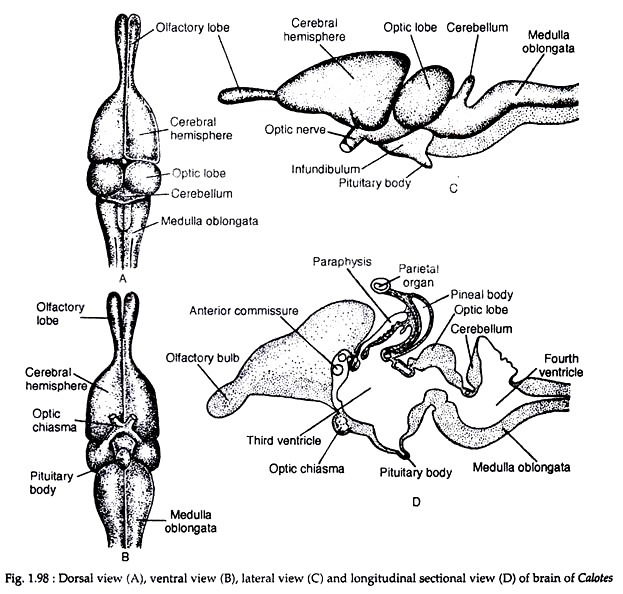
The midbrain consists of a pair of oval optic lobes or corpora bigemina which arise as projections of the dorsolateral walls. Ventral to the optic lobes, there are a pair of longitudinal bands or peduncles called crura cerebri, which connect the hindbrain to the midbrain. The hindbrain consists of narrow and non-convoluted metencephalon or cerebellum and a long myelencephalon or medulla oblongata, which continues posteriorly with the spinal cord.
Thickenings inside the different brain regions:
The roof of the cerebral hemispheres is thin but the ventrolateral walls are thick. The thick region is called corpus striatum. The roof of the cerebral hemispheres is called neopallium, because the grey matters are situated on the outer margin. The roof of diencephalon is thin, highly vascular and is called roof-plates (see Fig. 1.98D). The lateral walls are called thalami and the floor is called hypothalamus. Parietal and pineal bodies arise from the roof-plate.
The thalamus is thick. From the hypothalamus the infundibulum arises. The roof of the mesencephalon becomes thick to give rise to the optic lobes. Its floor is also thick and gives rise to the crura cerebri. The roof of the metencephalon is thin and non-nervous but the floor is thick. The myelencephalon is similar to metencephalon in regard to its floor and roof.
Ventricles:
ADVERTISEMENTS:
Internally, the brain bears cavities which are continuous to one another and to the spinal cord. The cavity is filled with cerebrospinal fluid. The cavities in the cerebral hemispheres are called lateral ventricles or first and second ventricles. The diencephalon contains the third ventricle while the fourth ventricle is situated in the medulla oblongata.
The two lateral ventricles are communicated to the third ventricle or diacoel by a small opening called foramen of Monro. The third and fourth ventricles or mesocoels are communicated with each other by a narrow passage called iter or aqueduct of Sylvius.
Commissures in Brain:
Two sides of the brain are connected at places by transverse bands of nerves called commissures.
The commissures are:
(1) Anterior commissure. It is a wide transverse tract of nerve fibres connecting the two corpora striata.
(2) Habenular commissure. It is anterior to epiphysis and situated between epithalamic ganglia.
(3) Aberrant commissure. It runs through the lamina terminalis and joins the olfactory lobes. The presence of aberrant commissure in the brain of Calotes is regarded as the most important diagnostic reptilian feature.
(4) Hippocampal commissure. The two hippocampi are connected by the hippocampal commissure.
(5) Posterior commissure. It connects the posterior part of the two optic thalami and is situated just at the junction of diencephalon and mesencephalon.
Spinal cord:
It is the posterior prolongation of the brain through the neural canal. Its walls are thick and the roof and the floor bear dorsal and ventral furrows. Externally the spinal cord is made up of white matter consisting of medullated nerve fibres and internally there is grey matter containing ganglionic cells and non-medullated fibres.
(b) Peripheral Nervous System:
The peripheral nervous system consists of nerve fibres which are either sensory or motor or mixed. The cranial and spinal nerves constitute this system.
Cranial nerves:
There are 12 pairs of cranial nerves in Calotes. The origin, distribution and biological nature of the first to tenth pairs of cranial nerves is exactly similar to that of Bufo. The origin and distribution of the ninth and tenth cranial nerves of Calotes is shown in Fig. 1.99. The eleventh and twelfth pairs of cranial nerves are the (a) spinal accessory, and (b) hypoglossal, respectively.

The spinal accessory (XI) originates from the posterior part of medulla oblongata as efferent fibres and joins with the vagus. Along its course, this nerve receives series of rootlets from the spinal cord.
The spinal accessory nerve supplies mostly the striated muscles of the pharynx, larynx and also the autonomic nervous system. It is a sensory nerve. The hypoglossal nerve (XII) is also a sensory nerve which originates from the medulla oblongata and innervates the muscles of the tongue and controls its movements during feeding.
Spinal nerves:
There are several pairs of spinal nerves that originate from the spinal cord and emerge out from it between the vertebrae. The spinal nerves are mixed type of nerves. They originate separately and independently but ultimately combine to form a single cord. They originate by a dorsal root which is sensory and a ventral root which is motor in nature. Each spinal nerve divides into four branches or rami.
These branches are:
(1) Dorsal branch, thin, short and supplies sense organs, glands and muscles.
(2) Ventral branch, long, thick and supplies hypo axial organs.
(3) Meningeal branch, small and goes back to supply the neural canal.
(4) Ramus communicans—which communicates with autonomic nervous system. Several spinal branches often meet together and form plexus.
(c) Autonomic Nervous System:
This system consists of a chain of complicated ganglia with receptor and effector nerves formed mostly by the offshoots from the ventral branches of the spinal nerves. The branches from these ganglia innervate muscles of heart, lungs, digestive system and glands which work continuously and are not controlled by will.
Autonomic nervous system comprises of two divisions of nerves which are structurally similar but functionally antagonistic.
These are:
(a) Sympathetic nerves having activating role.
(b) Parasympathetic nerves which have inhibitory action.
Sense Organs:
The sense organs are highly developed in Calotes:
(i) Olfactory organs:
The olfactory organs for smell in Calotes are the nasal chambers. The nasal chamber or passage extends between the external and internal nares. The anterior part of the nasal chamber is devoid of sensory cells while the posterior part bears many spindle-shaped cells connected to the twig of the olfactory nerve.
Highly segmented sensory epithelial cells cover the spindle shaped cells. To enlarge the surface of exposure, the epithelium of nasal wall is extended into a single shelf-like concha. Calotes has single unrolled concha one on either side.
(ii) Organ of sight:
The sense organ for sight consists of a pair of eyes which are lateral in position. The eyes are provided with eyelids and glands. The upper eyelid is small and less movable and the lower eyelid is large and more movable. The third eyelid or nictitating membrane is transparent. Harderian glands supply the third eyelid, while lacrimal glands make their first appearance in reptiles.
The wall of the globular eye is made up of several layers (Fig. 1.100A). The innermost layer bounding the posterior part of the eye is made up of retina. It is supplied with fine branches of nerves from the optic nerve. A vascular pigmented cushion-like projection arises from the blind spot region of retina. This projection is called pecten or ectodermal conus.
The layer outside the retina is called choroid which is made up of an inner pigmented layer and an outer vascular layer. Outside the choroid is situated the sclerotic layer which is protective in nature. The front of the eye is occupied by lens which is a clear ball derived from the skin (ectoderm). The iris is a pigmented and muscular ring in front of the lens.
The iris is a prolongation of the choroid. The aperture in this iris in front of the lens is called pupil. The front part of the eye is protected by cornea which is continuous with the sclera. The cornea is protected by conjunctiva, which is epidermal in nature. The lymph space between cornea and iris remains occupied by aqueous humor and the inner space between lens and retina remain filled with jelly-like vitreous humor.

(iii) Pineal Eye:
Majority of the reptiles possess a parietal organ on the dorsal side of the diencephalon and lying in the parietal foramen or just below it. The parietal organ develops as an outgrowth from the anterior part of the pineal body or directly from the cerebral roof. This organ often remains in close association with the skin.
The parietal organ has a vesicular appearance and the walls are composed of simple epithelium in Calotes, the parietal organ stays quite separate from the brain (Fig. 1.101). Because of the eye-like structure and on account of its connection with the roof of diencephalon or with the pineal body in many reptiles, the parietal organ is often referred as pineal or third eye.

Considerable controversies exist on the functional significance of the ‘pineal eye’. The skin over the top of the parietal organ, in some reptiles, becomes de-pigmented and assumes a cornea-like appearance. Despite eye-like appearance, the parietal organ has no trace of any visual function.
(iv) Organ of Jacobson:
An accessory structure, called organ of Jacobson or vomeronasal organ is associated with the olfactory organ of Calotes (Fig. 1.100B). It is a cavity derived embryonically from the olfactory cavity, lined with sensory epithelium and innervated by the vomeronasal division of the olfactory and also by the trigeminal nerves. It is a small sac-like structure with heavily pigmented walls supported by cartilages. An organ of Jacobson is present below each nasal cavity.
It is completely separated from the nasal cavity in adult. Paired ducts communicate with the mouth cavity. It acts as a chemoreceptor and it is highly developed in lizards and snakes. This sense organ appreciates scent particles introduced into it by its tongue tips. It has also been experimentally established that this organ in some reptiles helps in trailing prey and locating the opposite sex.
(v) Auditory sense organ:
This sense organ comprises of ears which perform two-fold functions—hearing and maintenance of equilibrium.
The ear of Calotes is made up of two parts:
(1) The middle ear, and
(2) The internal ear.
External ear is absent.
(1) Middle ear:
It is tubular cavity with its outer opening being covered by the tympanic membrane situated behind the eye. The membrane is slightly depressed and is perforated by very minute apertures. The inner end of the tube is covered by a very thin membrane. A bony rod-like structure called columella auris is situated inside the tube of the middle ear.
The role of this structure is to transmit the sound waves. The middle ear is communicated to the buccal cavity by a narrow but long tube called Eustachian tube. The middle ear communicates with the internal ear through a circular structure called fenestra ovalis.
(2) Internal ear:
The internal ear is situated in a bony capsule. The bony capsule has two fenestrae—fenestra ovalis and fenestra rotunda. The columella auris is fixed internally into the membrane which covers the fenestra ovalis. The space between the capsule and internal ear is filled in with perilymph. The internal ear is demarcated into an upper utriculus which is tubular and slightly bent and a lower sacculus which is sac-like.
Lagena or rudimentary cochlea is attached to the ventromedian side of the sacculus (Fig. 1.100C). The lagena is of simple type and contains the papilla basilaris, a special organ of hearing. From the utriculus arises a tubular structure called ductus endolymphaticus which opens beneath the duramater of brain.
There are three semicircular canals attached to the utriculus by both ends. Each semicircular canal is swollen at one end. The swollen part is called ampulla. The ampullae of anterior semicircular canal and horizontal semicircular canal are situated side by side while those of posterior semicircular canal and horizontal canal are situated at a distance.
Minute masses of CaCO3 present inside the ampullae help in maintaining balance. These pieces of CaCO3 are called statoliths or autoliths. The sacculus, utriculus and the three semicircular canals remain filled with a liquid called endolymph.