In this article we will discuss about the circulatory system of calotes.
Transportation of various substances within an organism is conducted by the circulatory system. The circulatory system consists of cardiovascular system and lymphatic system.
The cardiovascular system includes:
(a) The heart,
ADVERTISEMENTS:
(b) The blood,
(c) Arteries and
(d) Veins.
(a) Structure of Heart:
ADVERTISEMENTS:
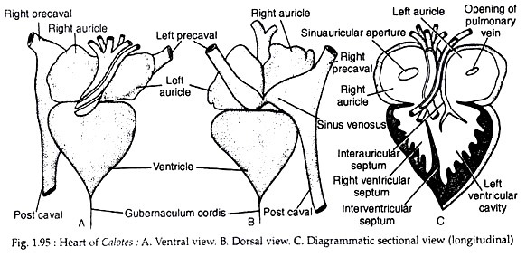
The heart of Calotes lies in the pleuroperitoneal cavity. The heart is covered by a thin and transparent pericardial membrane. The space between the heart and pericardium is filled with pericardial fluid. The heart is triangular in shape.
The auricular region is wider than the ventricular region (Fig. 1.95A). The sinus venosus is reduced and is disposed transversely and dorsal to the lower half of the auricles. It is thin-walled and is formed by the confluence of the venae cavae.
The right half of the sinus venosus is larger than its left counterpart and is formed by the confluence of right anterior vena cava (precaval vein) and posterior vena cava (postcaval vein). The left portion of the sinus venosus is composed mainly by the left anterior vena cava.
A constriction marks off the right and left parts of sinus venosus (Fig. 1.95B). The sinus venosus opens into the right auricle, near the region of the constriction, by a semicircular sinuauricular aperture. The aperture is provided with sinuauricular valves. The valves develop from the upper and lower margins of the aperture and the free end of the valves, which is slightly frilled, projects into the lumen of the right auricle.
ADVERTISEMENTS:
The right auricle is larger than the left auricle and appears darker than the left auricle. The wall of the right auricle is thick and its inner lining is thrown into a number of musculi pectinati which are projected within the lumen. The left auricle is smaller than the right auricle and it receives a common pulmonary vein.
The pulmonary aperture is circular in outline and is located on its dorsal wall close to the inter-auricular septum. The aperture is not provided with valves.
Internally, the left and right auricles are separated by a thin, muscular and non-perforated inter-auricular septum. The septum extends posteriorly for a short distance within the ventricle and bears at its posterior tip the auriculoventricular valves.
The ventricle is muscular, spongy and triangular in appearance. Its apex is directed caudally and bears a thin and white cord of tissue called gubernaculum cordis. It penetrates the pericardium and reaches the upper margin of liver. The thick-walled ventricle (Fig. 1.95C) is provided internally with an inter-ventricular septum which divides it incompletely into left and right halves.
The inner cavity of the ventricle has been arbitrarily divided into two regions, namely, cavum pulmonale situated in the right side, cavum arteriosum (on the left hand portion) by a muscular ridge. The ridge arises from the right ventral wall of the ventricle and runs to the dorsal side obliquely. Higher up, it becomes horizontally inclined.
It then runs obliquely again and takes a vertical course. It cuts off the aforesaid cavities. The left and right auriculoventricular apertures lie very close together being separated by the prolongation of the inter-auricular septum. The free lateral edge of the septum bears the auriculoventricular valves.
In the middle of the ventricle and close to the line of demarcation between the auricles and ventricle, there are three apertures from which the aortic arches arise. The inner wall of the ventricle is provided with thick interlacing muscles called columnae carnae. There are bunches of thread-like muscle fibres called chordae tendineae by which the valves remain attached to the columnae carnae (not shown in the figure).
The different compartments of the heart are intercommunicated by apertures having swing door-like flaps called the valves. These valves control the passage of blood and direct the flow in one direction.
ADVERTISEMENTS:
The valves present in the heart of Calotes are:
(a) A pair of leaf-like valves in the sinuauricular aperture.
(b) Sphincter muscle acting as valve in the opening of pulmonary vein into the left auricle,
(c) A pair of leaf-like valves formed by the bifurcation of the inter-auricular septum in the auriculo-ventricular aperture,
(d) Three semilunar valves in each orifice from which the arterial arches arise.
The walls of the heart are provided with three histological layers common to all blood vessels, i.e., tunica intima, tunica media and tunica adventitia from inner to the outside. Of these three layers, the tunica media is peculiar in having specialised cardiac muscles showing striations and branchings. The heart is supplied with the cardiac branch of 10th cranial nerve.
Mechanism of circulation through heart:
In Calotes, the circulatory circuit is double. These are pulmonary or lesser circulation and systemic or greater circulation. Pulmonary circulation is conducted by the pulmonary arteries which carry deoxygenated blood to the lungs. In the lungs, the blood becomes oxygenated and returns to the left auricle by the pulmonary vein.
The left auricle pours its content into the ventricle through the auriculoventricular aperture. In the greater circulation, deoxygenated blood returns to the sinus venosus by two precaval and one postcaval veins. The sinus venosus opens into the right auricle. The right auricle empties its content into the ventricle.
The ventricle sends blood for circulation into the different parts of the body through the systemic and pulmonary arches. The entry and exit of blood in the ventricle are so beautifully arranged that a major quantity of oxygenated blood is always forwarded to the brain region.
As the ventricle is incompletely divided, admixture of oxygenated and deoxygenated blood occurs. Thus, though the ventricle in Calotes is morphologically incompletely divided, there is a tendency for the physiological separation of the two types of blood, at least in two auricles completely and in the ventricle partially. From this point, the heart of Calotes is biologically more advanced than that of Bufo.
(b) Blood:
Blood of Calotes is red in colour and is made up of plasma and blood cells. The red blood corpuscles are biconvex, elliptical in outline and each bears an elliptical nucleus. The white blood corpuscles are irregular in outline, non-pigmented and each bears a spherical nucleus.
(c) Arterial System:
Of the six pairs of arterial arches joining the dorsal aorta to the ventral aorta during embryonic development of arteries, the third, fourth and sixth pairs persist in adult Calotes. As the ventricle in Calotes tends to divide into left and right ventricles by the development of incomplete inter-ventricular septum, the base of the ventral aorta splits into three parts, two of which remain in the right part of the ventricle and the third goes to the left part of the ventricle. Thus, from the ventricle of Calotes arise three aortic arches.
These arches are:
(1) One pulmonary aorta.
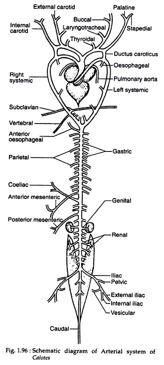
(2) Two systemic aortae, right and left (Fig. 1.96). These three aortae are wound around themselves at the source and undergo about one and half turns round each other. The aortae are covered at the base by a fibrous sheath and thus appear tubular.
Pulmonary aorta:
It arises independently from the right portion of the ventricle and soon splits into two branches, each entering into a lung. It carries deoxygenated blood.
Left systemic aorta:
This aorta originates independently from the right (left to pulmonary aorta) portion of the ventricle and moves forward for some distance. Then it curves round the heart and goes downwards to meet the right systemic aorta a little posterior to the apex of ventricle. It carries mostly the oxygenated blood.
From the left systemic arch four oesophageal arteries arise. The first of these arteries arises from near the point of insertion of the left ductus caroticus while the origin of the fourth one is very close to the point of union of the two systemics. Parietal arteries do not originate from the left systemic arch.
Right systemic aorta:
This important aorta emerges independently from the right ventral margin of the base of the ventricle and moves forward. It then curves to the right side of the heart. It meets the left systemic aorta posteriorly to form the dorsal aorta. It carries oxygenated blood. From the apex of the curvature of the right systemic aorta arises a single and common carotid artery which advances anteriorly and then splits into four arteries.
The inner pair of these four branches form the external carotid arteries while the outer pair form the internal carotid arteries. On both sides-prior to its division into left internal and external carotids and right internal and external carotids – one thyroid artery is given off from the common carotid.
The main branches from the right and left external carotids are the laryngotracheal artery (one from each) and three buccal arteries (from each). The internal carotids both left and right -bifurcate at their tips into inner palatine artery and outer stapedial artery. The external carotids and their branches supply blood to face and mouth, while the internal carotids and their branches supply blood to the brain.
The two internal carotids are connected to the systemic aorta of the corresponding sides by ductus caroticus. The ductus caroticus represents the remnant of embryonic radices between the third and fourth aortic arches.
From the right systemic aorta three oesophageal arteries are given out. The right systemic aorta before its meeting with the left systemic aorta gives rise to a subclavian artery which bifurcates into two and supplies the forelimbs.
From the right systemic aorta a vertebral artery also originates to send blood to the vertebral column. The point of origin of the vertebral artery is situated very close to the subclavian branch. Just before meeting its left counterpart, the right systemic arch gives a pair of parietal arteries.
The right and left systemic aortae unite a little behind the heart and give rise to the dorsal aorta which runs posteriorly and gives branches to visceral organs and posterior parts of the body. The following arteries originate from the dorsal aorta chronologically along the anteroposterior axis.
These are:
(a) Anterior oesophageal artery. It is single and originates from the ventral surface of the dorsal aorta.
(b) First pair of parietal arteries which plunge into the parieties of third thoracic vertebra.
(c) First and second pairs of gastric arteries, which supply the cardiac stomach.
(d) Second pair of parietal arteries.
(e) Third, fourth and fifth pairs of gastric arteries followed by third pair of parietal arteries.
(f) Sixth and seventh pairs of gastric arteries followed by fourth pair of parietal arteries.
(g) Eighth pair of gastric arteries followed by fifth and sixth pair of parietal arteries. It is to be noted that the number of gastric arteries varies from 4-8 pairs.
(h) Anterior mesenteric artery, which runs obliquely to supply the intestine.
(i) Coeliac artery which runs obliquely cranial, to supply the pyloric stomach. A splenic artery is given off by it to supply the spleen.
(j) Seventh and eighth pair of parietal arteries.
(k) Posterior mesenteric artery or hepato-intestinal artery. It arises from the right border of the dorsal aorta. It runs obliquely, and, on reaching the intestine, bifurcates into two — anterior and posterior. The anterior or hepatic supplies the gall-bladder while posterior or intestinal supplies the intestine.
(l) Ninth pair of parietal arteries.
(m) The right and left genital artery. The point of origin of the right one is a bit up than its counterpart on the left.
(n) Tenth and eleventh pairs of parietal arteries.
(o) Left and right renal arteries. There may be more than one pair.
(p) Twelfth and thirteen pairs of parietal arteries.
(q) One pair of iliac arteries. Each branch after its origin runs obliquely to the corresponding hind limb. From each iliac artery, a pelvic branch is given off to supply the pelvic girdle. Finally, each branch bifurcates into external and internal iliacs. From near the point of bifurcation a slender vesicular artery is given off by each branch.
(r) The dorsal aorta now enters into the tail as caudal artery.
(d) Venous System:
The deoxygenated blood from the different parts of the body is brought back to the heart by means of veins, except the pulmonary veins which carry oxygenated blood. The veins run parallel to the arteries and appear dark. These are positioned superficial to arteries.
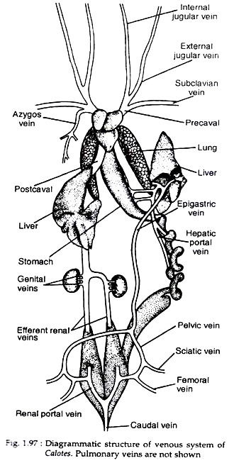
The central meeting arena of all veins in the body is the sinus venosus. Sinus venosus is a triangular structure and its two base angles receive left and right precavals while the apex receives a single median postcaval (Fig. 1.97). Each precaval vein has been formed by the union of three veins.
These are:
(1) The external jugular, which brings back blood from the floor of mouth and tongue.
(2) The internal jugular, which drains blood from the brain.
(3) The subclavian, which draws blood from the forelimb. The right precaval gets an azygos vein. The postcaval is constituted by the large median vein, which is formed by the union of right and left efferent renal veins emerging from the two kidneys. Genital veins join the left and right efferent renal veins before their union. A pair of stout but short hepatic veins joins the median postcaval before its entry into the sinus venosus.
A median caudal vein carries blood from the tail region. The caudal vein ultimately bifurcates into two veins which enter into the kidneys. Each vein gives rise to the renal portal vein to the kidney and pelvic vein which receives femoral and sciatic veins from the hind limb. The pelvic veins unite to form a median epigastric or anterior abdominal vein which, ultimately, opens into the left liver.
The anterior abdominal vein and the postcaval are free of each other except through the renal portals in the kidneys. The blood from the visceral organs, i.e., stomach, intestine, pancreas, etc., enters into the left lobe of the liver by a hepatic portal vein.
In Calotes both renal portal and hepatic portal systems are present. These systems have got many advantages and fulfill the demand for a second set of capillaries through which blood must flow. The organisms having such a portal system are always provided with double supplies of blood, arterial and venous.
The pulmonary venous circuit comprises of pulmonary veins. From each lung, two pulmonary veins carry blood to the heart. Of these veins, one comes out from the anterior part while the other comes from the posterior part of lung. Near the left auricle all these four branches unite and then open into the left auricle. The pulmonary veins bring oxygenated blood to the heart from the lungs.
The lymphatic system is highly developed. The main lymphatic trunk becomes divided and enters into the precaval veins. Lymph hearts are present.


