In this article we will discuss about the development stage of chick embryo, fertilized eggs are procured from recognised poultry farm and incubated in the laboratory.
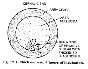
1. Chick: M. 4 Hours of Incubation:
1. Four hours after incubation of the egg shows differentiation of the blastodisc into area pellucida and area opaca. (Fig. 7).
2. One quadrant of area pelluciada becomes thickened, which marks the future caudal end of embryo.
ADVERTISEMENTS:
3. After 7 to 8 hours, the thickening becomes more elongated and represented start of primitive streak.
2. Chick: W.M. 16 Hours Embryo:
Comments:
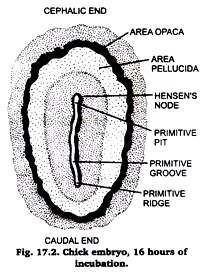
1. 16 hours after incubation the primitive streak becomes so distinct that embryos are characterized as being in primitive streak stage. (Fig. 8).
2. In fixed and stained slide, w.m. is composed of central furrow, called as primitive groove lined by thickened primitive ridges.
ADVERTISEMENTS:
3. At the cephalic end of the primitive streak, closely-packed cells form thickened area, called as Hensen’s node. Part of area pellucida adjacent to the primitive streak shows increased thickness and forms embryonic elliptical shape.
4. Area pellucida assumes elliptical shape.
5. Elongated primitive streak represents long axis of future embryonic body.
ADVERTISEMENTS:
6. Caudal end of the streak is that which lies close to the area opaca.
3. W.M. 18 Hours Chick Embryo:
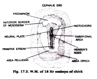
1. It is a W.M. of 18 hours stage of chick embryo.
2. At this stage the dark peripheral area opaca and central translucent area pellucida are distinctly visible.
3. In the anterior part is present the pro-amnion, which is a small and comparatively more translucent region of area pellucida and is characterised by the absence of mesoderm.
4. In the middle of area pellucida, in the posterior half, runs a primitive streak having a primitive groove through its centre. The primitive groove is being bound by primitive folds.
5. In the anterior half of area pellucida, in the middle, runs a neural groove bound by neural folds.
6. The primitive streak and neural groove is separated by a thickening-the Hensen’s node having a small depression in the centre-the Hensen’s pit.
7. The primitive streak gives rise to an out-growth, the notochord immediately below the primitive groove.
4. W.M. of 21 Hours Chick Embryo:
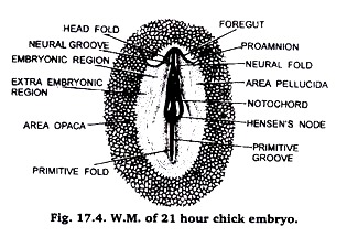
1. It is a W.M. of 21 hours chick embryo.
ADVERTISEMENTS:
2. At this stage the dark peripheral area opaca and central translucent and colourless area pellucida are distinctly visible.
3. In the anterior part are present the pro-anmnion, which is a small and comparatively more translucent region of area pellucida and is characterised by the absence of mesoderm.
4. In the middle of area pellucida, in the posterior half, runs a primitive streak having a primitive groove through its centre. The primitive groove is being bound by primitive folds.
5. In the anterior half of area pellucida, in the middle, runs a neural groove bound by neural folds.
6. The primitive streak and neural groove are separated by a thickening, the Hensen’s nod having a small depression in the centre of the Hensen’s pit.
7. The primitive streak gives rise to a small outgrowth, the notochord immediately below the primitive groove and to mesoderm on either side.
8. At this stage embryonic and extra ambryonic regions have also become distinguished in the area pellucida.
9. In the anterior most part the ectoderm has given rise to head fold, which is a pocket-like extension of neural folds.
10. With the ectoderm the underlying endoderm is also transformed into a pocket-like structure the -foregut.
11. The proambion is comparatively reduced in size.
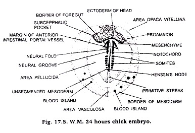
5. W.M. of 24 Hours or 4 Pairs of Somites Stage of Chick Embryo:
1. It is a W.M. of 24 hours 4 pairs of somites stage of chick embryo.
2. At this stage the dark peripheral area opaca and central translucent and colourless area pellucida are distinctly visible.
3. In the anterior part is present the proamnion, which is a small and comparatively more translucent region of area pellucida and is characterised by the absence of mesoderm.
4. In the middle of area pellucida, in its posterior half runs a primitive streak with a primitive groove in its centre. The primitive groove is bound by primitive folds.
5. In the anterior half of area pellucida, in the middle, runs the neural groove bound by neural folds.
6. The primitive streak and neural groove are separated by Hensen’s node having a small depression in the centre-the Hensen’s pit.
7. Immediately below the primitive groove the primitive streak gives rise to a small out- growth, the notochord and on either side to mesoderm.
8. In the area pellucida embryonic and extra embryonic regions also become distinguished.
9. In the anterior- most part the ectoderm has given rise to head fold, which is a pocket-like extension of neural folds. The underlying endoderm is also transformed into a pocket-like foregut. The proamnion is greatly reduced.
10. In front of Hensen’s node the mesoderm of embryonic area differentiated into 3-4 pairs of mesodermal somites.
11. The neural canal, in the region of head fold, gives rise to forebrain.
12. The foregut extends on either side into an amino-cardiac vesicle.
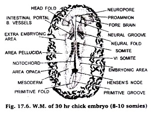
6. W.M. of 30 Hours of 8-10 Pairs of Somites Chick Embryo:
1. It is W.M. of 30 hours of chick embryo or 8-10 pairs of somite stage of chick embryo.
2. At this stage the dark peripheral area opaca and central translucent and clourless area pellucida are distinctly visible.
3. In the anterior part is present the proamnion, which is a small and comparatively more translucent region of area pellucida and is characterised by the absence of mesoderm.
4. In the middle of area pellucida, in the posterior half, runs a primitive streak with a primitive groove running through its centre. The primitive groove is bound by primitive folds.
5. In the anterior half of area pellucida, in the middle, runs the neural groove bound by neural folds.
6. The primitive streak and neural groove are separated by Hensen’s node having a small Hensen’s pit in the centre.
7. Immediately below primitive groove the primitive streak gives rise to the notochord and on either side to mesoderm.
8. At this stage embryonic and extra embryonic regions have also become distinguished in the area pellucida.
9. In the anterior-most part, the ectoderm has given rise to head fold which is a pocket like extention of neural folds. The underlying endoderm has transformed into pocket like foregut. The proamnion is reduced.
10. The mesoderm, in front of Hensen’s node, has given rise to 8-10 pairs of somites.
11. In the region of head fold the anterior part of neural canal has given rise to a distinct fore brain.
12. The foregut and cardiac vesicles are sufficiently developed.
13. The extra embryonic area has grown in size.
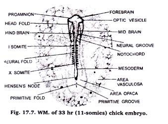
7. W. M. of 33 Hour Chick Embryo of 11-12 Pairs Somites:
1. It is W.M. of 33 hours chick embryo.
2. At this stage the dark peripheral area opaca and central translucent area pellucida are not distinctly visible.
3. The primitive streak has been comparatively reduced because of great lengthening of neural canal and neural folds.
4. The extra embryonic area has grown in size.
5. The mesoderm, in front of Hensen’s node, has given rise to 11-12 pairs of somites.
6. The foregut and cardiac vesicles are sufficiently developed.
7. The brain is differentiated into fore brain, mid- brain and hind brain.
8. The area opaca has changed into area vasculosa.
9. Proamnion has disappeared.
10. Anterior omphalomesenteric vein has developed.
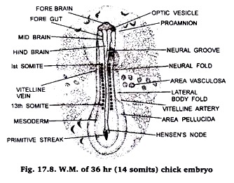
8. W.M. of Chick Embryo of 13-14 Pairs Somites or 36 Hours:
1. It is W.M. of 36 hours chick embryo.
2. At this stage the dark peripheral area opaca and central translucent and colourless area pellucida are not visible.
3. The extra embryonic area has grown in size.
4. The primitive streak is comparatively reduced because of great lengthening of neural canal and neural folds. The notochord has extended from behind the brain up to the end of body.
5. The mesoderm, in front of Hensen’s node, has given rise to 13-14 pairs of somites.
6. The brain is differentiated into fore brain, mid brain and hind brain.
7. In the fore brain region optic vesicles and in the hind brain region optic vesicles have developed.
8. The area opaca has changed into area vasculosa.
9. Proamnion has disappeared.
10. Anterior omphalomesentric vein and vitelline artery have developed.
11. The cardiac vesicle has given rise to heart.
9. W.M. of 48 Hours Chick Embryo of 26-28 Pairs of Somites:
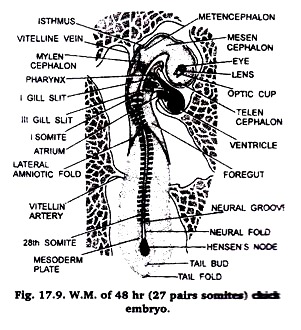
1. It is W.M. of 48 hours chick embryo.
2. At this stage the area opaca and area pellucida are not visible.
3. The extra embryonic area has grown in size.
4. Primitive streak has disappeared.
5. The mesoderm, in front of Hensen’s node, has given rise to 26-28 pairs of somites.
6. The brain has differentiated into telencephalon, prosencephalon, mesencephalon, metancephalon and mylencephalon.
7. The heart has been differentiated into ventricle and atrium. Sinus venosus and truncusarteriosus have also started developing.
8. The eye has been differentiated into optic cup and lens and optic vesicle has also developed sufficiently.
9. The head region has curved on right side due to cranial flexion.
10. Three pharyngeal gill-slits have also been differentiated.
11. Behind Hensen’ node a tail bud has also developed.
12. Lateral amniotic folds, anterior omphalomesentric vein and vitelline artery have appeared.
10. W.M. of 72 Hours or 36 Pairs of Somites Stage of Chick Embryo:
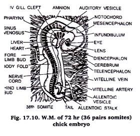
1. It is W.M. of 72 hours chick embryo.
2. At this stage area opaca and area pellucida are not visible.
3. The extra embryonic area has grown in size.
4. Primitive streak has disappeared.
5. The mesoderm, in front of Hensen’s node, has given rise to 36 pairs of somites.
6. The brain has differentiated into telencephalon, mesencephalon, metancephalon and mylencephalon.
7. The heart has been differentiated into ventricle and atrium.
8. The eye has differentiated into optic cup and lens and optic vesicle has also developed sufficiently.
9. The head region has bent on right side due to cranial flexion.
10. Four pairs of gill-slits have been differentiated.
11. Tail bud is greatly developed and has given rise to allentoic stalk and tail.
12. Lateral amniotic folds, vitelline artery and anterior omphalomesentric vein have developed.
13. In the middle region a pair of fore limb buds and in front of tail a pair of hind limb buds have developed, which will give rise to fore and hind limbs.
14. Olfactory pit, visceral arches, amnion, allantois and amniotic cavity have also developed.
11. W.M. of 96 Hours Chick Embryo:
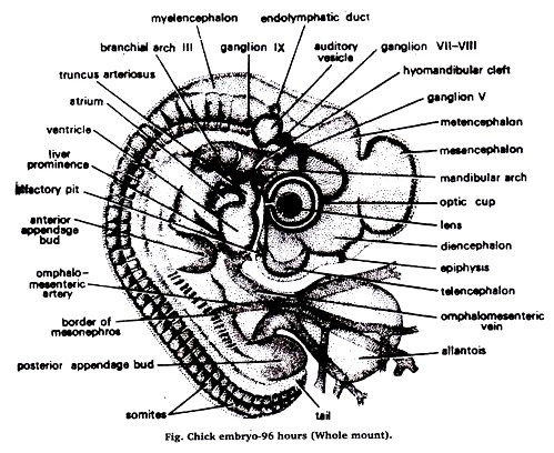
1. In the chick embryo of 96-hours of incubation, the entire body has been turned through 90 degree and the embryo lies with its left side on the yolk.
2. At the end of 96 hours the body folds have undercut the embryo so that it remains attached to the yolk only by a slender stalk.
3. The yolk salk soon become enclogated, allowing the embryo to become first straight in the mid-dorsal region and then convex dorsally.
4. The progressive increase in the cranial, cervical, dorsal and caudal flexures results in the bending of the embryo on itself so that its originally straight long axis becomes C-shaped and its head and tail lie close together.
5. Optic cup shows the more developed lens.
6. Endo-lymphatic duct arises from the auditory vesicle.
7. Visceral arches have become very much thickened.
8. Appendage buds increase rapidly in size and become elongated.
9. The number of somites increases to 41 pairs.
10. Allantois has also appeared.
11. Omphalomesenteric artery and omphalomesenteric vein are also developed.
