In this article we will discuss about:- 1. Structure of Hydra 2. Body Wall of Hydra 3. Cell Elements 4. Locomotion 5. Nutrition 6. Respiration and Excretion 7. Nervous System 8. Reproduction 9. Regeneration.
Contents:
- Structure of Hydra
- Body Wall of Hydra
- Cell Elements of Hydra
- Locomotion in Hydra
- Nutrition in Hydra
- Respiration and Excretion in Hydra
- Nervous System of Hydra
- Reproduction in Hydra
- Regeneration in Hydra
1. Structure of Hydra:
Hydra belongs to the class Hydrozoa, Phylum cnidaria.
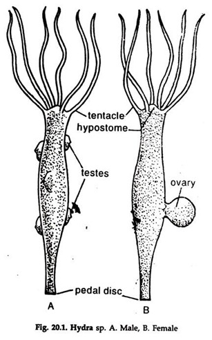
1. The body is more or less cylindrical (Fig. 20.1) and one end remains attached to a submerged object, wood or stone.
2. The attached end is known as the proximal end, the point of attachment is the pedal disc.
3. The distal or the opposite end is provided with 6 or 8 tentacles radiating in all directions and arranged in a circle.
4. Hypostome is a small, elevated cone surrounded by the tentacles.
5. The mouth is round and situated at the centre of the hypostome.
ADVERTISEMENTS:
6. The mouth communicates the coelenteron or the gastro-vascular cavity to the exterior.
7. The whole surface of the body, except the pedal disc, is provided with a large number of stinging cells or cnidocytes (cnidoblasts);
i. The cnidocytes are crowded in the hypostome and the tentacles.
8. One or more buds, the tubular outgrowths, similar to the structure of the body are often present, as lateral outgrowths of the body.
ADVERTISEMENTS:
9. Small, conical outgrowths, male or female gonads, also called testis or ovary, are present on the body.
10. Indian Hydra is unisexual.
2. Body Wall of Hydra:
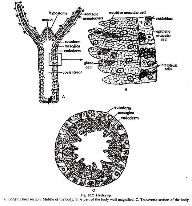
Hydra is the simplest fresh water metazoa. It is the smallest and solitary cnidarian polyp. The wall of the body, tentacles and buds are made up of two layers of cells (Fig. 20.2), the outer ectoderm and the inner endoderm separated by a thin layer of non-cellular gelatinous mesogloea. The ectodermal layer is protective, receptive and reproductive and the endodermal layer is mainly nutritive.
3. Cell Elements of Hydra:
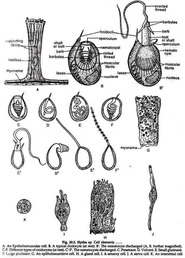
Of the different types of cells some are restricted to ectoderm, some to the endoderm and some are common to both ectoderm and endoderm. (Fig. 20.3)
Cells Confined to Ecotoderm:
a. Epitheliomuscular Cells:
These are large elongated cells with oval nucleus. The free ends are smooth with rounded corners and being placed side by side form a continuous surface. Two basal strands, opposite each other, are present in each cell.
ADVERTISEMENTS:
A myoneme extends through them at the base of the cell. By the contraction of these processes the animal is shortened and can bend in various directions. The cells of the pedal disc can send pseudo- podia by which hydra can move (Fig. 20. 3A).
b. Cnidocytes:
Cnidocytes (Cnidoblasts) are weapons of offence and food capture. It has the following components (Fig. 20.3B-F).
a. A tough, ovoid, fluid-filled capsule, the nematocyst, invaginated at the anterior end and continuing as a long, hollow thread, bearing minute barbs at the base.
b. A layer of specially contractile cytoplasm with a large nucleus.
c. A delicate protoplasmic process, the cnidocil or trigger hair at the anterior end, projecting beyond the surface.
d. Contact of the cnidocil with some small animals causes sudden contraction of the cytoplasm, the nematocyst is everted and shot out with a great force.
Usually four types of cnidocytes are present and distributed in a characteristic regional pattern. They are most concentrated in the tentacles and anterior region, become less numerous towards the base and absent in the basal disc.
Types of Cnidocytes:
i. Penetrant or stenotele:
Large, about 16 µm, spherical; the long thread remains coiled in a transverse plane. Three pairs of barbs and rows of small thorns or nettles are present at the base (Fig. 20.3C). The discharged thread pierces the animal body and the fluid hypo-toxin, protein in nature, in the nematocyst, has a numbing effect on the animals.
ii. Volvent or desmoneme:
Pear-shaped, about 9 µm in size; thread short, in the form of a single loop (Fig. 20.3D). The thread coils round the prey after discharge.
iii. Glutinant steroline or atrichus isorhize:
Size about 7 µm; thread unarmed and straight (Fig 20.3E).
iv. Glutinant streptoline or holotrichous:
Oval, about 9 µm. in size; thread long with three or four transverse coils and bears small nettles (Fig. 20.3F). The thread coils round the prey after discharge.
The penetrant and volvent types help in capture of preys while the streptoline and steroline glutinant types help both in food capture and locomotion.
Cnidocytes once used become non-functional and cast off.
c. Reproductive cells:
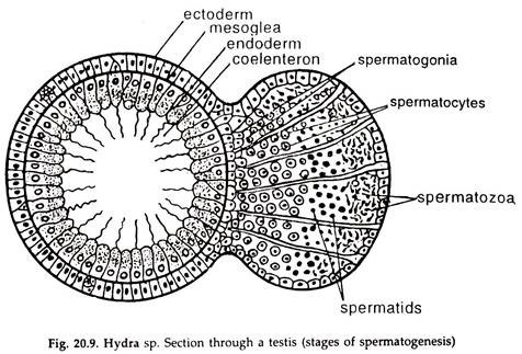
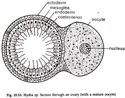
Hydra is dioecious or monoecious. Undifferentiated male and female germ cells are present in the ectodermal layer which, in the breeding season, multiply to form the gonads, testis or ovary (Figs. 20.9, 20.10).
Cells Confined to Endoderm:
i. Epithelionutritive or Nutritive- muscular cells:
Large cells (Fig. 20.3G) with one or more vacuoles and a prominent nucleus. Two flagella are present at the free tip of some of them. The basal end is provided with two contractile extensions containing a myoneme. The extensions take a circular course next to mesogloea. The Hydra elongates when these processes contract. These cells carry out nutritive function.
ii. Mucous cells:
Modified gland cells situated in the oral gastro dermis and possess flagella. Their secretion helps in the lubrication of food for easy swallowing.
iii. Albumen cells:
Elongated cells with narrow bases and may or may not possess flagella. They supply albumen to the developing ovum.
iv. Storage cells:
Modified gland cells.
Cells Common to Ectoderm and Endoderm:
a. Gland cells:
Abundant in tentacles, oral region and pedal disc. In Hydra, the pedal disc is thickly populated (beset) with glandulo-muscular cells, and they have muscular basal extensions (Fig. 20.3H). In the gastro dermis, they secrete digestive ferments.
b. Sensory cells:
Elongated cells found in the tentacular and the oral region. Their bases are continuous into one or more fine fibrils passing into the general nerve plexuses (Fig. 20.31).
c. Nerve cells:
Commonly known as ganglion cells, which conduct functional impulses (20.3 J).
d. Interstitial cells:
Situated between the narrow bases of the epithelial cells and are considered as persistent undifferentiated embryonic cells. They are capable of forming cnidocytes, transforming into sex cells as well as other types of cells. They are also supposed to take part in building and repairing processes. (Fig.20.3 K).
4. Locomotion in Hydra:
Hydro usually remains attached to a submerged object or the substratum under water with the basal disc and remains in an upright position. The tentacles are in constant movement in search of food and also in response to certain abiotic factors.
Locomotion in Hydra (Figs. 20. 4. -20.6) is Effected in the Following Ways:
A. Gliding:
The hydra glides over the substratum with the amoeboid movement of the cells of the basal disc, without any change in the erect body position.
B. Floating:
Occasionally the cells of the basal disc secrete gas bubble, which makes the body light and enables the hydra to float on the water surface. In that state, it is carried away by wind or water current. The tentacles often help in swimming.
C. Looping:
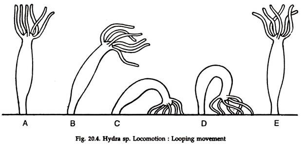
It is the most common type of locomotion in hydra. It resembles the looping of a caterpillar (Fig. 20.4). In the normal erect position the body elongates, followed by bending of the distal part and the tentacles are attached to the substratum with the help of glutinant cnidocyles.
The basal disc is released, the proximal end drawn forward and the basal disc reattached to the substratum, close to the tentacles, followed by assuming the erect position.
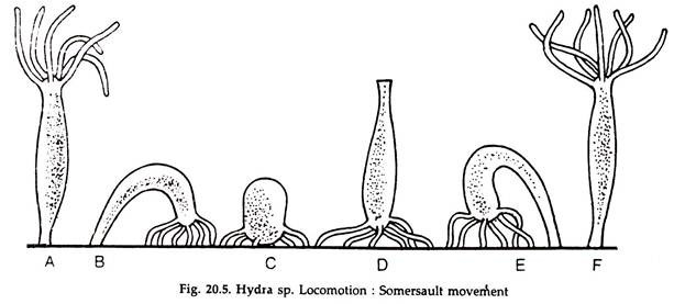
D. Somersaulting:
The first phase of somersaulting resembles looping. The erect body extends and bends, the tentacles are attached to the substratum forming a loop. The basal disc is freed, the animal stands on the tentacles and the body contracts to form a small knob.
This is followed by extension of the body and bending by which the basal disc is placed on the substratum in a forward position, forming a loop. The tentacles are released and the animal assumes an upright position (Fig. 20.5).
By repeating the process, hydra moves to a distant place. This is the normal mode of locomotion in hydra.
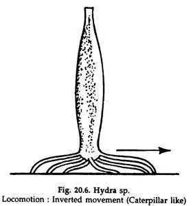
E. Inverted:
The tentacles are fixed to the substratum, the basal disc is up and the hydra is in an inverted position (Fig. 20.6). By alternate contraction and relaxation of the tentacles, the hydra moves forward slowly.
F. Climbing:
Hydra relaxes the tentacles and attaches them with aquatic vegetation or any object. The basal disc is released and tentacles contract, pulling the body to a new position.
5. Nutrition in Hydra:
Hydra can digest proteins, fats, some carbohydrates, but not starch.
1. Small annelids, crustaceans, insect larvae are chief food of hydra.
2. The nematocysts discharge in contact with the food animal and the toxin injected into the body of the victim paralyses the prey.
3. The paralyzed prey is wrapped about by tentacles and gradually drawn towards the mouth which opens widely to receive it.
4. Usually, hydra swallows only living prey and it engulfs only those animals which contain glutathione.
5. Glutathione is present in the tissue fluids of most animals and is necessary to evoke the feeding reactions in hydra.
6. The engulfed materials are covered by mucous secreted by the mucous gland cells in the first part of coelenteron.
7. Proteolytic enzyme is secreted in the enteron from the gland cells.
8. In the alkaline medium extracellular digestion of protein occurs.
9. Some endodermal cells produce pseudopodia and engulf the partly digested food into food vacuoles.
10. The rest of the digestion take place in food vacuoles, which is intracellular digestion.
11. The digested food is absorbed by the endoderm cells and then transferred to ectoderm and distributed to all parts of the body.
12. Undigested food materials are ejected through the mouth on contraction of the body.
6. Respiration and Excretion in Hydra:
Hydra has no blood, no blood vessels, nor any organ of respiration and excretion. As the body wall is thin and the gastro vascular cavity usually circulated with water, most of the cells of the body remain freely exposed to the surrounding water.
The exchange of oxygen and carbon dioxide gases—necessary for respiration and excretion of nitrogenous wastes (chiefly ammonia) occurs directly by diffusion through the cell membranes as in protozoans.
7. Nervous System of Hydra:
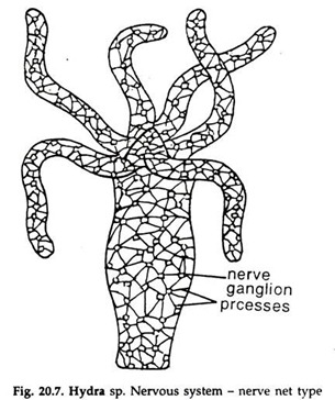
Hydra is the first metazoan in which a nervous system is met with. A network formed by the nerve ganglia and the processes of the neurons constitute the nervous system (Fig 20.7) and termed as nerve net system.
8. Reproduction in Hydra:
Hydra reproduces both by asexual and sexual means:
I. Asexual Reproduction:
Both budding and fission are present, the first one is most common.
A. Budding:
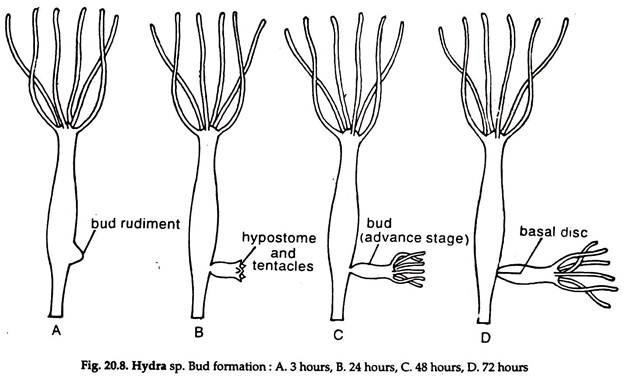
1. It occurs in summer in well-fed and healthy hydra when food supply is abundant (Fig. 20.8).
2. The interstitial cells in the ectoderm, near the base or about the midway of the cylinder, multiply rapidly forming a slight swelling and appear as a hollow projection— the bud, containing ectoderm, mesoglea and endoderm with an out-pocket of coelenteron.
3. Gradually the bud elongates, a mouth opens at its free distal end and a ring of blunt tentacles sprouts at the base of the hypostome.
4. A little hydra with mouth, tentacles, stalk and gastro vascular cavity, remaining still continuous with the parental one is formed.
5. Gradually the base region constricts, the young hydra separates from the parent body and starts an independent life.
6. Sometimes several lends may develop at a time in a hydra.
B. Fission:
Occasionally both transverse and longitudinal fission occur in hydra, although it is not a normal mode of reproduction. Accidental breakdown, or for regaining normal shape from atypical condition, such as persistence of buds, two heads, two discs, etc. leads to -such fission.
II. Sexual Reproduction:
1. Most hydras are dioecious but some species are monoecious (hermaphrodite) in which sperms mature before the eggs, and self-fertilization is prevented.
2. Sexual reproduction usually occurs in winter and spring months.
3. In some species (Hydra oligactis) male is smaller and bears one to eight testes, while the larger female bears one or two ovaries.
4. Sex reversal has also been observed where in one season a hydra develops ovaries but in the next season it develops testes.
5. In hermaphrodite species usually testes develop towards the distal end and the ovaries towards the basal disc.
6. Lowering of temperature and increase in free carbon dioxide in water promote the formation of gonads in hydra.
Development of testes:
1. A testes is a slight conical elevation, arising from ectodermal interstitial cells and having a number of elongate cysts.
2. At the base of a cyst is the primordial germ cell (interstitial cells), which divide repeatedly by mitosis to form a variable number of spermatogonia, which grow into spermatocytes (Fig.20.9).
3. The spermatocytes undergo meiotic division forming spermatids which develop into spermatozoa.
4. A mature spermatozoon is minute, possessing three parts—a swollen head containing nucleus, a narrow mid-piece, and a long slender tail.
5. The mature sperms make their way through the rupture of outer ectodermal wall.
6. After release the sperms usually remain viable for 24 to 48 hrs.
Development of ovary:
1. The development of ovary is similar to that of testis. The interstitial cells multiply to form the germ mother cells or oogonia.
2. One centrally located cell, the oocyte, becomes larger and amoeboid with a large nucleus and yolk materials.
3. Subsequently, it undergoes a meiotic division to produce one ovum and two polar bodies.
4. One ovary usually contains a single ovum and remains attached to the parent by a broad base. Surrounding the ovum a gelatinous sheath is present (Fig. 20.10).
Fertilization and Development in Hydra:
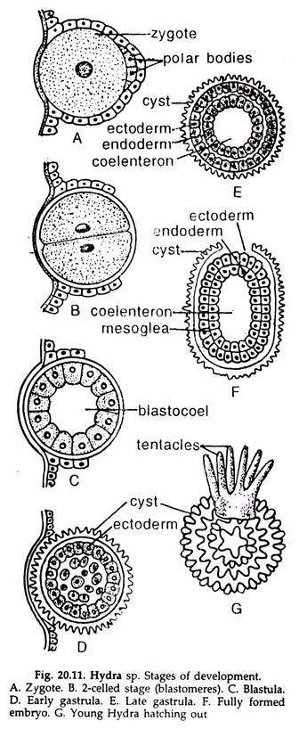
a. Several sperms penetrate the gelatinous sheath but only one can successfully enter the ovum and fertilize it to form a zygote (Fig. 20.11).
b. The zygote cleaves immediately. The cleavage is holoblastic and results in a hollow spherical ball, the blastula or coeloblastula. The cavity inside it is the blastocoel.
c. Gradual development leads to a solid gastrula or stereo gastrula.
d. The inner mass of cells derived from the blastomeres form the endoderm. A new cavity called archenteron or gastrocoel gradually develops in this solid mass.
e. The ectodermal cells secrete a two layered protective shell called cyst or theca around gastrula. The outer layer of shell is thick, horny or spiny but the inner layer is a thin gelatinous membrane.
f. The gastrula is shed from the body before the formation of shell. It either sinks in the mud at the bottom or sticks to aquatic plants.
g. As the cyst can withstand drying or freezing, it remains unchanged until the next spring or suitable time.
h. Within the cyst the embryo elongates, a circlet of tentacles develop at one end surrounding the month.
i. As the embryo grows the outer shell softens and breaks open, releasing the young Hydra.
j. The young hydra now fixes itself to a substratum and gradually assumes adult characteristics.
9. Regeneration in Hydra:
i. Regeneration is the ability to restore lost or worn out parts of the body. The capacity to replace or regenerate parts lost by injury or otherwise is allied to growth after fragmentation.
ii. Young animals and species low in the evolutionary scale usually have greater regenerative power than the older or higher animals.
iii. Regeneration power in hydra was reported first by Trembly (1744). Hydra, if cut into several pieces, each will regenerate the lost parts and develop a whole new individual.
iv. The body parts usually retain their polarity in original, with oral end developing tentacles and aboral end, basal disc.
v. If the head of hydra is split into two and the parts are slightly separated, it results into a Y-shaped hydra or two-headed individual, having two mouths and two sets of tentacles.
vi. Brien (1953) and others found that in hydra there is a growth zone just below the base of the tentacles. The interstitial cells give rise to new body cells of all types. As the new cells are formed, other cells are pushed towards the- tentacles or the basal disc where the old cells are shed. All the cells may be renewed within 45 days.
vii. If the internal cells of the growth zone are destroyed by X-rays, the hydra lives only for a few days.
