In this article we will discuss about the silk glands of silkworms.
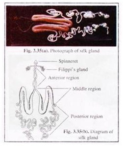
The silk of silkworms is secreted by a pair of labial gland, known as silk glands. The silk glands lie ventral to the alimentary canal. In full grown larvae, these occupy most of the body cavity. The silk glands are tubular in shape with different diameters in different regions. Each gland has 3 distinct regions [Fig. 3.35(a & b)].
(1) Posterior region:
ADVERTISEMENTS:
Blunt, highly folded tubular posterior regions of both glands remain attached to tracheal bushes of silkworm. This part secretes fibroin as fibrinogen which converted to fibroin upon extrusion.
(2) Middle region:
Most prominent and widest part of silk gland. It remains folded in a W-shaped structure and thus has 3 limbs — posterior, middle and anterior limbs. The posterior arm secretes sericin-I. It gets surrounded by serecin-II secreted from the middle limb.
This sericin again gets surrounded by sericin- III secreted from the anterior limb. The middle region of silk gland also acts as the reservoir of fibroin where the later gets mature during the storage period.
ADVERTISEMENTS:
(3) Anterior region:
The thin anterior region of silk gland has no secretory role and only transports the assembled silk to the spinneret.
Spinneret:
It is a projection of the median part of the labium, which draws the silk out in the form of fine filament. The secreted silk comes out as a thread or filament as it passes through silk press which resembles a typical salivary pump. The two filaments coming out of two sides are called brins. The sericin (gum) layer of the two brins then bind together into a single filament or bave.
ADVERTISEMENTS:
Histologically the entire gland has 3 layers:
(1) The outer tunica propia with uniform thickness;
(2) The middle glandular layer with gland cells which increase in size during later instar stages of larval development and
(3) The inner tunica intima: It has varying thickness.
In the anterior region of the gland, this layer is very thick and is shed at each moult. In other regions of silk gland, it is thin and not shed at each moult.
Filippi’s gland or Lyonnet’s gland:
In the head region of the larvae, a pair of glands is situated which open into the anterior part of silk gland near its opening into the spinneret. It is thought that these glands contribute some waxy materials to the silk thread or lubricate the passage of silk while coming out.
