In this article we will discuss about the circulatory system of toad.
The circulatory system includes two systems: the blood vascular system (cardio vascular system) and the lymphatic system. Circulatory system is the internal transport mechanism by which nutritive materials, hormones, waste products, carbon dioxide and oxygen are conveyed to the different parts of the body.
The cardiovascular system of toad is well-developed. This system is composed of three main components: blood, heart and blood vessels.
(i) Blood:
ADVERTISEMENTS:
Blood is the main circulatory fluid. It consists of a straw-coloured fluid called blood plasma and different blood corpuscles suspended in the plasma. The plasma is a watery liquid and contains many mineral salts, food, wastes and hormones. The corpuscles are of three types.
These are red blood cells (RBC or erythrocytes), white blood cells (WBC or leucocytes) and blood platelets (or thrombocytes). The erythrocytes are oval, biconvex and nucleated cells ranging from 15 to 20 micron in size. They contain the red coloured iron-containing protein, the haemoglobin.
The leucocytes are colourless, nucleated and amoeboid cells. They are phagocytes and destroy bacteria and thus protect the animal from the invading microbes. The thrombocytes or blood platelets are small spindle-shaped nucleated cells and play an important role in the process of coagulation of blood.
(ii) Heart:
ADVERTISEMENTS:
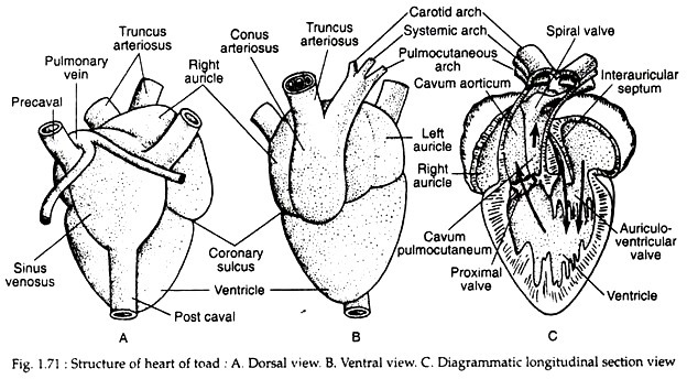
Heart is the central pumping organ in the cardiovascular system (Fig. 1.71). In toad, it is a pear-shaped muscular structure situated in the anterior part of the body cavity. It remains enclosed by a transparent protective membrane called pericardium. The space between heart and the pericardium is called pericardial cavity.
The heart of toad is composed of the receiving parts and forwarding parts. The receiving parts include two auricles and a sinus venosus. The ventricle and the conus arteriosus are the forwarding parts of the heart. Inter-communicating apertures and valves between the different parts of the heart are present.
The auricles are two in number and are of unequal size. Both the auricles are sharply marked off from the ventricle by a narrow constriction called coronary sulcus. The left auricle is smaller than the right auricle. The two auricles are completely separated by inter-auricular septum.
ADVERTISEMENTS:
The sinus venosus is a triangular thin- walled chamber formed by the union of three major veins — two precavals and one post-caval (Fig. 1.71A). It is situated on the dorsal side of the right auricle. It communicates with the right auricle through a sinuauricular aperture which is guarded by sinuauricular valves.
Deoxygenated blood, collected in the sinus venosus from the precaval and postcaval veins, enters into the right auricle but the back-flow is prevented by the sinuauricular valves. The left auricle receives oxygenated blood through a small opening of the common pulmonary vein. This aperture is also guarded by valves.
The right and left auricles communicate to the ventricle by a common auriculo-ventricular aperture. This aperture is guarded by membranous valves known as auriculo- ventricular valves. The free ends of the valves are attached with the wall of the ventricle by fine thread-like chordate tendineae. The auriculo-ventricular valves allow one-way traffic of blood from the auricles to Ventricle.
The ventricle is a highly muscular chamber. It is more or less conical in shape. Its cavity is greatly reduced by a large number of interlacing muscles called columnae carnae. The conus arteriosus is a stout tubular body situated on the ventral side of the heart (Fig. 1.71B). It is continued anteriorly into the truncus arteriosus.
The truncus arteriosus is not to be considered as the part of heart. It is the basal stem of the three main arteries. It remains uncovered by the pericardium and lacks cardiac muscles. A set of pocket-like semilunar valves demarcate the truncus from the conus. A similar set of three semilunar valves guard the opening between the conus and the ventricle. These valves prevent back flow of blood from conus to the ventricle.
A twisted longitudinal spiral valve divides the cavity of the conus into two channels. The left channel is designated the cavum pulmocutaneum and the right one is the cavum aorticum (Fig. 1.71C). The deoxygenated blood passes through the cavum pulmocutaneum and the oxygenated blood travels through the cavum aorticum by the manipulation of the spiral valve.
Mechanism of circulation through the heart:
Periodic contraction (systole) and relaxation (diastole) is an innate property of heart. During distole, the sinus venosus receives deoxygenated blood from the two precavals and one postcaval veins. The left auricle is also simultaneously filled up with oxygenated blood from the lungs, carried through the common pulmonary vein.
Just with the onset of systole, the sinus venosus contracts and the deoxygenated blood is rushed into the right auricle through sinuauricular aperture. So both the auricles simultaneously get filled up with blood — the left one with oxygenated blood and the right with deoxygenated blood.
ADVERTISEMENTS:
After the auricles get filled up, the auricular systole starts. Two auricles contract simultaneously and drive the contents into the ventricle through the auriculoventricular aperture. Consequently two types of blood enter into the cavity of the ventricle where admixture of the two types takes place.
Due to the spongy nature of the ventricular cavity, a major quantity of the deoxygenated blood is kept in the right side; the left side contains mostly the oxygenated blood while the middle part contains mixed type of blood. No separation of the oxygenated blood actually occurs in the ventricular cavity. As the skin sub-serves respiratory function, the right auricle also receives oxygenated blood.
Next the ventricle starts contraction. The back flow of blood into the auricles is prevented by the auriculoventricular valves. The blood from the ventricular cavity finds its way through the conus. As the conus arises from the right side, a large quantity of deoxygenated blood from the ventricle enters first.
This blood is now conveyed through the cavum pulmocutaneum by the spiral valve. From the cavum pulmocutaneum blood goes to pulmocutaneous arteries for oxygenation. The deoxygenated blood is aerated in lungs and brought back to left auricle through common pulmonary vein and thus completes the pulmonary circuit.
With the enhancement of contraction force of the ventricle, the mixed blood from the middle region of the ventricle is pushed into the systemic arches through the cavum aorticum. Lastly, the pressure exerted by the carotid labyrinth is overcome and mostly the oxygenated blood from the left side of the ventricle is forcefully pumped into the carotid arches. It is to be noted that the spiral valve directs the entry of blood into different arches.
(iii) Blood Vessels:
The arteries and veins are the blood vessels through which the blood circulates. These vessels are different in structure and function. The arteries break up into finer branches, the arterioles which finally end into a network of capillaries. These capillaries again reunite to form small venules. The venules unite to form the veins.
Histologically, a typical blood vessel is composed of three distinct layers. The outermost covering is made up of fibrous connective tissue known as tunica adventitia. The middle layer is composed largely of involuntary muscles called tunica media. The innermost layer is made up of endothelium and elastic fibres and is called tunica interna.
Arterial System:
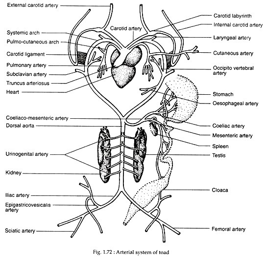
The arteries and their branches constitute the arterial system (Fig. 1.73). The truncus arteriosus divides into two main branches, each of which again splits up into three arches – the anterior carotid arch, the middle systemic arch and the posteriormost pulmocutaneous arch.
Embryonic arrangement of the arterial arches:
In the embryonic state, six pairs of arterial arches are present. Of these paired arches, the first and second pairs disappear in an adult. The third pair are converted into the carotid arches. The fourth pair are transformed into the systemic arches. The fifth pair disappear and the sixth pair persist as the pulmocutaneous arches in adult.
Carotid arches:
There are two carotid arches in toad. Each carotid arch proceeds forward and outward. It soon divides into an outer branch called internal carotid artery supplying blood to the brain, the meninges and into an inner branch called external carotid artery which supplies blood to the outer side of the head, thoracic musculature, tongue and the buccal cavity.
Just at the point of bifurcation and towards the base of internal carotid artery, there is a small swelling known as carotid labyrinth (or carotid gland). This gland is derived from the remnants of gill and connecting blood vessel between the first afferent and efferent branchial arteries. The inner cavity of the carotid labyrinth contains a network of small vessels and forms a spongy structure.
Though this structure is a receptor, it is physiologically connected with the regulation of the blood pressure. It controls blood pressure in the internal carotid artery. The internal carotid artery, after its origin, takes a backward course and comes very close to the systemic arch and is tied with it by fibrous carotid ligament.
Systemic arches:
There are two systemic arches in toad. Each systemic arch takes the median position and sweeps outward to surround the oesophagus. It then comes to the dorsal side and joins with its fellow of the opposite end to form the dorsal aorta.
The following branches arise from each systemic arch:
(1) A laryngeal artery which is very short and supplies the laryngotracheal chamber.
(2) An occipitovertebral artery which gives branches to the pharynx, back of the head, vertebral column and spinal cord.
(3) A stout subclavian artery supplies blood to the shoulder and forelimb.
All these branches exhibit bilateral symmetry but the left systemic arch gives off an additional branch called oesophageal artery which supplies blood to the oesophagus. This branch is not present on the right side. The dorsal aorta occupies the mid-dorsal position and is situated ventral to the vertebral column and ends posteriorly into two iliac arteries.
The dorsal aorta gives off the following branches anteroposteriorly:
(1) A stout coeliacomesenteric artery which emerges out just from the origin of the dorsal aorta. The artery immediately breaks up into a coeliac branch to supply blood to the stomach, liver, gall bladder and pancreas and a mesenteric artery supplying blood to the mesenteries, intestine, cloaca and spleen.
(2) Four or five pairs of renal arteries which supply to the kidneys. From the anterior-renal arteries, additional branches arise for the gonads. These small branches are called genital arteries. The renal arteries and genital arteries are collectively known as urinogenital arteries.
(3) The iliac arteries are the last branches of the dorsal aorta. From each iliac artery an epigastricovisceralis artery is given off to supply the urinary bladder and the ventral body wall. The iliac artery then enters into the hind limb and divides into femoral and sciatic arteries.
Pulmocutaneous arches:
These are the shortest and the hindermost arches which carry deoxygenated blood to the lungs and skin. Each main arch enters into lung as pulmonary artery and a very slender branch from it goes to the skin as cutaneous artery.
Venous System:
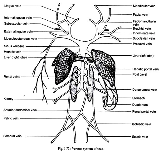
The veins and their branches constitute the venous system (Fig. 1.73).
The venous system of toad can be described under three headings:
(1) Systemic,
(2) Portal, and
(3) Pulmonary.
Systemic veins:
Three large veins or venae cavae which open into the sinus venosus represent the systemic veins. The systemic veins carry deoxygenated blood from almost all parts of the body except lungs. The anterior two venae cavae are known as left and right precavals and the single posterior one is called postcaval. Each precaval vein is formed by the union of three branches.
These are:
(i) External jugular vein,
(ii) Innominate vein, and
(iii) Subclavian vein.
The external jugular vein is formed by two veins, a lingual carrying blood from the tongue and a faciomandibular from the snout and jaws. The innominate vein is also formed by the union of two veins, an internal jugular bringing blood from the head and a subscapular from the back of shoulder.
The subclavian vein is similarly formed by two veins, a brachial vein bringing blood from the forelimb and a musculocutaneous vein from the muscles and skin. As the skin acts as an accessory respiratory organ, the musculocutaneous vein brings oxygenated blood.
The postcaval vein is formed by four or five pairs of renal veins which receive blood from the kidneys. Blood from reproductive organs is also poured into renal veins by the genital veins. The postcaval vein then ascends to enter into the sinus venosus.
Portal veins:
A portal vein has its origin in capillaries and ends in capillaries. The blood from the portal vein returns to the heart through an intermediate organ. The hepatic and renal portal systems are two portal systems in toad. The capillaries from the gut unite to form a hepatic portal vein, which again breaks up into capillaries in liver.
The capillaries from the posterior part of the body unite to form two renal portal veins which, in turn, break up into capillaries in the kidneys on their way to the heart.
Hepatic portal system:
This system comprises of hepatic portal vein and the anterior abdominal vein or epigastric vein. Hepatic portal vein carries blood from stomach, intestine, pancreas and spleen. The main vessels receive the anterior abdominal vein under the liver and enter into the substance of the liver.
The anterior abdominal vein is formed by the union of two pelvic veins in the mid-ventral line. The pelvic vein arises as an offshoot of the femoral vein. On its anterior, the abdominal vein receives small branches from the urinary bladder and the ventral body wall.
Renal portal system:
The blood from the hind limbs is carried by the femoral and sciatic veins. Each femoral vein on entering the body cavity gives off a pelvic vein. The main trunk of the femoral vein receives the sciatic vein above the level of the pelvic vein to form the renal portal vein.
The renal portal vein proceeds by the side of the corresponding kidney and enters into it to break up into capillaries. Each renal portal vein receives two or three dorsolumbar veins carrying blood from the body wall.
Pulmonary veins:
Two pulmonary veins arises from two lungs and unite and opens into the left atrium of the heart (not shown in figure). Oxygenated blood from the lungs comes to the heart. So, pulmonary veins carry oxygenated blood, which is the exception among veins.
Lymphatic System:
While circulating through the body, the blood does not come directly into the cells. During the transit of blood through capillaries plasma exudes from the capillary wall into the intercellular spaces as the lymph. It is a colourless fluid with few leucocytes. The lymph from the intercellular spaces is collected by lymphatic vessels which in turn lead into lymph sacs.
Some of the lymph vessels become contractile and are known as lymph hearts which pump back the lymph into the veins. A pair of lymph-hearts opening into femoral vein is present near the urostyle and another pair are situated below the scapulae which open into subscapular veins. Some openings of small dimension in the peritoneum communicate with the lymphatic vessels or lymphatics.


