In this article we will discuss about:- 1. Meaning of Ribosome 2. Types of Ribosome 3. Structure 4. Chemical Composition 5. Biogenesis 6. Functions.
Contents:
- Meaning of Ribosome
- Types of Ribosome
- Structure of Ribosomes
- Chemical Composition of Ribosomes
- Biogenesis of Ribosomes
- Functions of Ribosome
1. Meaning of Ribosome:
Ribosomes are small, rounded, dense particles of ribonucleoprotein. They occur either freely in the cytoplasm and the matrix of mitochondria and chloroplast or remain attached with the membranes of the endoplasmic reticulum and nucleus.
ADVERTISEMENTS:
They are the sites of protein synthesis in both prokaryotic and eukaryotic cells. The cells in which active protein synthesis takes place (e.g., pancreatic cells, hepatic parenchymal cells, plasma cells, thyroid cells etc.), the ribosomes remain attached with the membranes of RER.
Cells which synthesize specific proteins for the intracellular utilization and storage like erythroblasts, skin etc. contain large number of free ribosomes in the cytoplasm. Ribosomes are usually designated according to their rates of sedimentation. The sedimentation co-efficient is expressed in the Svedberg unit, or S unit (S is related with the size and molecular weight of the ribosomal particles).
2. Types of Ribosome:
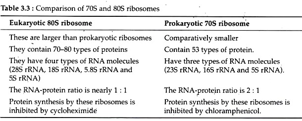
According to the size and sedimentation co-efficient two types of ribosome have been recognized (Table 3.3).
i. 70S Ribosomes:
They are found in prokaryotic cells of blue green algae and bacteria. They are smaller in size and have the sedimentation co-efficient of 70S and molecular weight 27 x 106 Daltons.
ii. 80S Ribosomes:
They occur in eukaryotic cells of plants and animals. They have the sedimentation co-efficient of 80S and molecular weight 40 x 106 Daltons. The sedimentation value of the ribosomes of mitochondria and chloroplasts vary in different phyla, e.g., 77S in mitochondria of fungi, 60S in mitochondria of mammals. The ribosomes of chloroplasts are of 70S type.
ADVERTISEMENTS:
ADVERTISEMENTS:
3. Structure of Ribosomes:
The general structures of prokaryotic and eukaryotic ribosomes are similar. Each ribosome is porous, hydrated and composed of two subunits. One subunit is large in size and has a dome-like shape, while the other subunit is smaller in size and occurring above the larger subunit, forming a cap-like structure.
In 70S ribosome the large subunit is 50S and the smaller is 30S. The 80S ribosomes also consist of two subunits, viz., 60S and 40S. Both subunits remain separated by a narrow cleft. The two subunits remain united with each other due to high concentration (0.001 M) of the Mg++.
When the concentration of Mg++ reduces in the matrix, both ribosomal subunits get separated. In bacterial cells, the two subunits are found to occur freely in the cytoplasm and they unite only during protein synthesis.
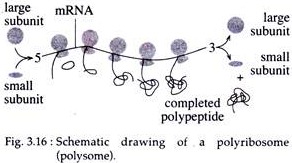
During protein synthesis in both prokaryotes and eukaryotes, many ribosomes bind to an individual mRNA molecule. The ribosomes are spaced as close as 80 nucleotides apart along a single messenger RNA molecule. The structures are called polyribosomes or polysomes (Fig. 3.16).
Ultra-structure of Ribosomes:
Ultra- structure of ribosomes has been studied more extensively in prokaryotes than eukaryotes. Negative staining of ribosomes has led to better understanding of the fine-structure of these organelles.
A. Models of 70S Ribosome:
ADVERTISEMENTS:
i. Quasi-symmetrical Model (Stoffler and Wittmann, 1970):
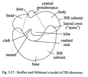
According to this model, the smaller 30S subunit of prokaryote ribosomes has a bipartite structure with an elongated, slightly bent pro-late shape. A transverse cleft divides the subunit into two parts; a smaller head and a larger body, giving it the appearance of a ‘telephone receiver or embryo’.
Under electron microscope, the frontal view of larger subunit shows three protuberances (Fig. 3.17) arising from a rounded base; the central being the most prominent and often gives the larger subunit the appearance of an arm chair (the rounded base forms the vaulted seat, the central protuberance forms the back and the lateral protuberances the arm of the chair) or a maple leaf.
During association, the frontal face of 30S subunit faces the vaulted seat of the 50S sub-unit. The long axis of 30S subunit is oriented transversely to the central protuberance of the 50S subunit. So a tunnel is formed between the hollow of the small subunit and vaulted seat of the large subunit (Fig. 3.17).
ii. Asymmetrical Model (J. A. Lake, 1981):
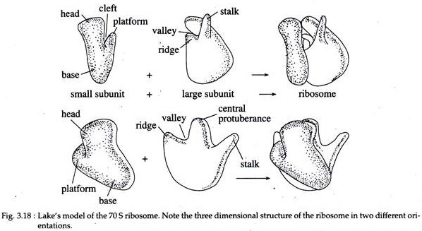
According to this model, the smaller subunit has a head, a base and a platform (Fig. 3.18). The platform separates the head from the base by a cleft.
The cleft is an important functional region the site of codonanticodon interaction and part of binding site for the initiation factor of protein synthesis. On the other hand, the large subunit consists of a ridge, a central protuberance and a stalk. The first two are separated with the help of a valley.
B. Three Dimensional Model of 80S Ribosome:
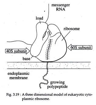
The cytoplasmic ribosomes of eukaryotes are remarkably similar in morphology to those of the prokaryotes. Like the 30S subunit of prokaryotes, the 40S subunit of eukaryotes is divided into head and base segments by a transverse groove (Fig. 3.19). The 60S subunit is rounded in shape, although its one side is flattened and it becomes confluent with the small subunit during the formation of the monomer.
A ribosome contains three binding sitet for RNA molecules. One for mRNA and two for tRNAs; one of the last two is called the peptidyl-tRNA binding site, or P-site and the other is called the aminoacyl-tRNA binding site, or A-site.
4. Chemical Composition of Ribosomes:
Chemically the ribosomes are composed of RNA and proteins. More than half of the weight of ribosome is RNA. The 70S ribosomes contain three types of rRNA, viz., 23S rRNA, 16S rRNA and 5S rRNA.
The 23S and 5S rRNAs are present in the larger 50S sub- unit, while the 16S rRNA occurs in the smaller 30S ribosomal subunit. The 23S rRNA consists of 2,900 nucleotides, 16S rRNA 1,540 nucleotides and 5S rRNA 120 nucleotides (Fig. 3.20).
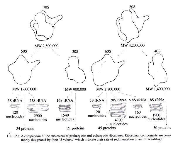 The 80S ribosomes contain four types of rRNA, viz., 28S rRNA with 4,700 nucleotides, 5.8S rRNA with 160 nucleotides and 5S rRNA with 120 nucleotides in the larger 60S subunit and the smaller subunit of 40S contains 18S rRNA with 1,900 nucleotides (Fig. 3.20).
The 55S ribosomes of mammalian mitochondria lack 5S rRNA but contain 21S and 12S rRNAs of which 21S rRNA occurs in the larger or 35S subunit, while 12S rRNA occurs in the smaller or 25S subunit.
About 60 percent of the rRNA is helical or double stranded and contains paired bases. These regions are due to hairpin loops between complementary regions of the linear molecules. In ribosome, the RNA is exposed at the surface of the ribosomal subunits and the protein is assumed to be in the interior, in relation to non-helical part of the RNA.
The 70S ribosome of E.coli is composed of about 55 types of ribosomal proteins, of which, 21 types have been isolated from the 30S subunit (designated in order of decreasing size S 1 through S 21), and 34 proteins (LI through L34) from the 50S subunit (Fig. 3.20).
In E.coli, all the proteins of the two sub- units are different, with the exception of one that is present in both subunits (S 20 and L 26). So, the total number of proteins in one ribosome is 54. Most of them are rich in basic amino acids. The smaller subunit (40S) of eukaryotic ribosome is composed of approximately 30 proteins and the larger subunit (60S) contains about 45 proteins.
In addition to rRNA and proteins, ribosomes also contains some divalent metallic ions, such as Mg++, Ca++‘and Mn++. However, eukaryotic ribosomes do not differ functionally from those in prokaryotes; they perform the same functions by the same set of chemical reactions. Eukaryotic ribosomes are able to translate bacterial mRNAs efficiently, provided that a ‘cap’ is added enzymatically.
5. Biogenesis of Ribosomes:
Ribosomes are not self-replicating particles. The synthesis of various components of ribosomes such as rRNAs and proteins are under genetic control. In E. coli the synthesis of ribosomal proteins is controlled at the translational level.
Some of the ribosomal proteins that bind directly to rRNA can also bind to similar structure in their own mRNA. In eukaryotes, the biogenesis of ribosomes is the result of the co-ordinated assembly of several molecular products that converge upon the nucleolus.
The 18S, 5.8S and 28S RNAs are synthesized as part of a much longer precursor molecule in the nucleolus, 5S RNA is synthesized on the chromosomes outside the nucleolus, and ribosomal proteins are synthesized in the cytoplasm. All these components migrate to the nucleolus, where they are assembled into ribosomal subunits and transported to the cytoplasm.
6. Functions of Ribosome:
Ribosomes take part in protein synthesis. Two or more ribosomes simultaneously engaged in protein synthesis on the same mRNA-strand forming polyribosomes. Interaction of the tRNA-amino acid complex with mRNA brings about translation of the genetic code, coordinated by the ribosomes (see translation).
During protein synthesis, ribosomes play a protective function. The mRNA strand which passes between two subunits of the ribosome is protected from the action of nucleases. Similarly the nascent polypeptide chains passing through the tunnel or channel of the larger subunit of ribosome are protected against the action of protein digesting enzymes.
