In this article we will discuss about the axial skeleton in vertebrates with the help of suitable diagrams.
Skull:
The skeletal framework of a vertebrate head is called a skull. A lamprey has a skull consisting of a braincase and cartilages of the tongue. A shark has a skull made of a braincase and isolated upper and lower jaw bars.
But commonly in animals ranging from fishes to mammals, the skull has a brain case to which the upper jaws are welded by a series of bones, the lower jaw is not included.
However, the skull of vertebrates is derived from three different embryonic components:
ADVERTISEMENTS:
1. Chondrocranium:
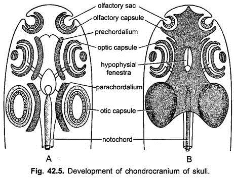
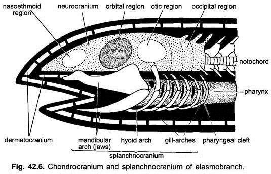
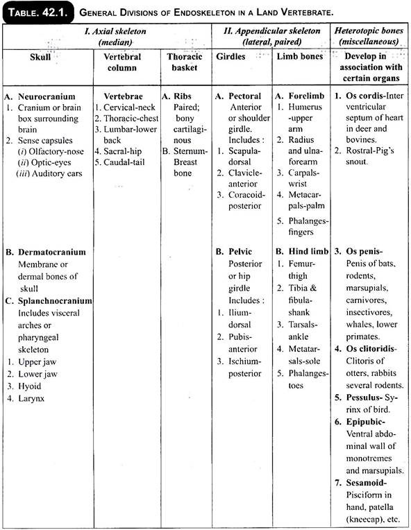
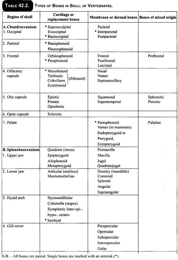
It includes cartilaginous brain case or neurocranium and cartilaginous capsules of olfactory, optic, and otic sense organs. The chondrocranium is replaced by bones in most vertebrates.
2. Splanchnocranium:
It is derived from the visceral or pharyngeal skeleton which is cartilaginous but becomes largely replaced or invested by bones in higher forms. It provides support to the gills and forms the jaws and suspensorium in gnathostomes.
ADVERTISEMENTS:
3. Dermatocranium:
It consists of dermal bones which become attached to the chondrocranium and splancfinocranium in bony fishes and tetrapoda. A dermatocranium is absent in cyclostomes, elasmobranchs, and a few higher fishes because the entire skull is cartilaginous.
Development of Chondrocranium:
After the formation of central nervous system and notochord in the embryo, the mesenchyme cells form a membranous covering around the brain and the anterior part of the notochord. Cartilage is formed in this membrane. It gives rise to a pair of cartilaginous plates, the parachordals lying below the midbrain and hindbrain and anterior part of the notochord.
ADVERTISEMENTS:
In front of the parachordals forms a pair of curved cartilaginous rods, the trabeculae or prechordals lying below the anterior part of the brain. At the same time cartilaginous capsules arise around the developing sense organs. They are a pair of olfactory capsules around the organs of smell, a pair of optic capsules around the eyes, and a pair of auditory or otic capsules around the auditory organs (internal ears).
The parachordals grow larger and fuse in the middle line forming a basilar plate leaving a small opening or basicranial fenestra. The two prechordals or trabeculae grow towards each other and fuse in the midline to form an ethmoid plate.
The ethmoid plate and the basilar plate grow towards each other and fuse to form a single basal plate forming the floor of the brain. A large opening, the hypophyseal fenestra is present in the basal plate, which lodges the pituitary gland.
The olfactory and otic capsules join the basal plate to form a cartilaginous chondrocranium. The optic capsules are sometime cartilaginous but more usually are fibrous, and never fuse with the chondrocranium but remain free and form the sclerotic of the eye, thus, they permit mobility of eyes.
The floor of the brain, thus, formed by the basal plate begins to grow upwards on the sides. In elasmobranchs the side walls grow and meet above the brain to enclose it in a brain- box or neurocranium. A few openings are left uncovered for the cranial nerves and blood vessels.
But in most other vertebrates there is no roof of cartilage, only the posterior or occipital region gets roofed over by cartilage and the rest of the brainbox has only a dorsal membrane. Later on membrane or dermal bones form the roof of brain. The formation of chondrocranium leaves a large opening or foramen magnum behind through which the spinal cord emerges.
Development of Splanchnocranium:
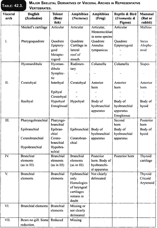
A visceral skeleton is formed partly from the neural crest cells and from splanchnic mesoderm around the pharynx between gill-clefts for their support. It consists of series of paired visceral bars (usually seven pairs) of cartilage which become united with one another ventrally by an unpaired cartilage to form visceral arches.
The visceral arches are horse-shoe-shaped and encircle the pharynx all around except dorsally. Typically there are seven visceral arches in fishes, though this number varies form four to nine in different groups. The first visceral arch is the mandibular arch, the second is the hyoid arch, the remaining ones are branchial arches because they generally support gills in lower aquatic vertebrates.
ADVERTISEMENTS:
In all vertebrates, except Agnatha, the mandibular arch forms jaws for supporting the mouth. This arch on either side is divided into a dorsal palatopterygoquadrate cartilage and a ventral Meckel’s cartilage. The palatopterygoquadrate or palatoquadrate forms the upper jaw, while the Meckel’s cartilage forms the lower jaw.
The second or hyoid arch on either side gives off a dorsal hyomandibular cartilage to support and connect jaws to chondrocranium beneath auditory region and a ventral cartilage which forms the hyoid apparatus which gives support to the tongue.
The remaining or branchial arches form support for the gills or larynx. The branchial arches support the gills in fishes and in tetrapoda these are much reduced, and form the hyoid apparatus and cartilages of the larynx.
Development of Dermatocranium:
The formation of the skull stops at the cartilaginous stage in cyclostomes and Chodrichthyes, but from bony fishes upwards cartilage bones and dermal or membrane bones become incorporated with the skull.
The dermal bones constitute the dermatocranium; they appear in the head region of bony fishes as scales. These dermal scales are actually parts of the exoskeleton which sink inwards and fuse with the roof of the chondrocranium to complete a protective envelope around the brain.
The dermal bones may remain independent of the rest of the skull so that they can be removed easily by boiling, or they may become fused with cartilage and cartilage bones so that all distinction between replacing and dermal bones is lost in the adult.
Jaw Suspension or Suspensoria:
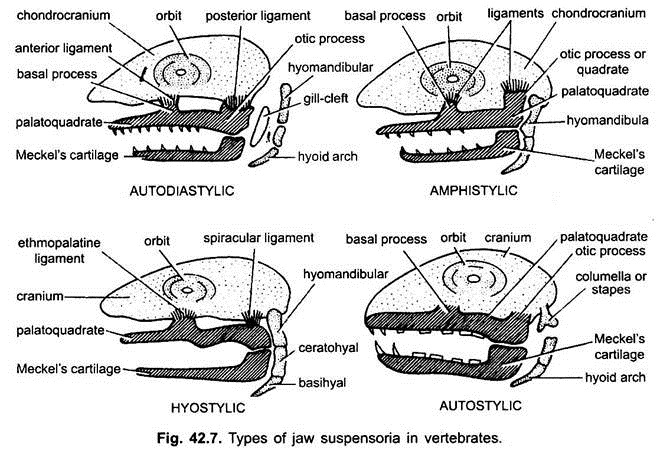
The method by which the upper and lower jaws are suspended or attached from the chondrocranium is known as jaw suspension or suspensorium. Amongst the visceral arches, the first (mandibular) arch consists of a dorsal palatopterygoquadrate bar forming the upper jaw, and ventral Meckel’s cartilage forms the lower jaw.
The second (hyoid) arch consists of a dorsal hyomandibular supporting and suspending the jaws with the cranium, and a ventral hyoid. The remaining visceral arches support the gills and are, hence, called branchial arches. Thus, splanchnocranium forms the jaws and suspends them with the chondrocranium.
Suspensoria are of five types:
1. Autodiastylic:
The jaws are attached by ligaments (anterior and posterior) to the chondrocranium. The hyoid arch does not support the jaws but remain completely free as the posterior branchial arches. The gill-cleft in front of the hyoid arch does not form a spiracle but forms a complete gill, e.g., early bony fishes (acanthodians).
2. Amphistylic:
The upper jaw (mandibular arch) has basal and otic processes which are attached by ligaments to the chondrocranium. Besides this, the hyomandibular of the hyoid arch is also attached to the chondrocranium. At the other end both jaws are suspended from it. Thus, it is a double suspension in which both the mandibular and hyoid arches are attached to the chondrocranium. This type of suspensorium is found in Crossopterygii and in some primitive sharks Heptanchus and Hexanchus.
3. Hyostylic:
The upper jaw is (palatoquadrate) is loosely articulated with the cranium by anterior ethmopalatine ligament and posterior spiracular ligament. Both jaws are suspended from the hyomandibular which is attached to the otic region of the skull. Thus, only hyoid arch binds both the jaws with the cranium and, hence, it is called hyostylic. It is found in most elasmobranchs and bony fishes. These fishes are able to swallow large preys.
In fishes the autodiastylic suspension is the most primitive, the amphistylic condition was derived from it, while the hyostylic condition is the most recent having been arrived independently.
4. Autostylic:
The upper jaw (palatoquadrate) is completely fused by its processes to the bony skull and the articular of lower jaw is suspended from the quadrate of the upper jaw. The hyomandibular do not take part in suspensorium and modified into columella or stapes of the middle ear.
Some authorities use the term autosystylic for autostylic. It is found in extinct placoderms, chimaera, lung fishes and tetrapods. i.e, amphibians, reptiles and birds. In these, the quadrate of the upper jaw articulates with the articular of the lower jaw.
The autostylic suspension is divided into three varieties:
(a) Holostylic:
The upper jaw is fused to the skull and the lower jaw is suspended from it. The hyoid arch is complete and not attached to the skull, e.g., Holocephali (chimaera).
(b) Monimostylic:
In many tetrapoda, except mammals, hyomandibular forms columella (middle ear bone) and the articular of lower jaw articulates with the quadrate of the upper jaw. The quadrate becomes an immovable part of the skull.
(c) Streptostylic:
In lizards, snakes and birds the articulation is between quadrate and articular, but the quadrate is not firmly fused with the skull and is movable at both ends. This autostylic suspension is distinguished as streptostylic.
5. Crainostylic:
The upper jaw is fused with the cranium in its entire length. Hyomandibular forms the stapes of middle ear bone. The quadrate and articular also modified into malleus and incus respectively. Thus, squamosal of the skull and dentary of lower jaw articulate with each other and both are dermal bones. It is found in mammals. Some consider it as modification of autostylic type.
Vertebral Column:
Notochord:
The primitive axial skeleton is a notochord present in all chordate embryos. It is formed from chordamesoderm cells. It is a stiff rod below the nerve cord and above the alimentary canal running from the infundibulum to the hind end of the body.
At first the cells of the notochord are packed closely but later they fuse together and become vacuolated, except a layer of peripheral cells. The notochord is enclosed in an inner and outer elastic fibrous sheath of connective tissue called elastica interna and elastica externa respectively.
In protochordates (Amphioxus) and cyclostomes (lamprey) the axial skeleton is primarily the notochord, which persists throughout life of the animal. In hagfishes small cartilaginous elements are present in the caudal region, and in lampreys two pairs of cartilaginous elements occur on either side of the spinal cord in each body segment.
They do not meet above to form a neural arch. In some of lower bony fishes (sturgeon), the notochord persists unchanged and, although neural and haemal arches are present, a centrum is lacking. In chimaeras and lungfishes, cartilage begin to invade the notochordal sheath.
In most fishes centrum is well developed. In fishes and higher animals, notochord is later surrounded by cartilaginous or bony rings called vertebrae. Notochord is practically obliterated in tetrapods.
The vertebral column is made of a metameric series of vertebrae extending longitudinally from the skull to the tip of the tail.
Vertebrae:
Vertebrae of various animals are different, even in the same vertebral column there are differences, but all vertebrae conform to a basic plan. Generally fish vertebrae are divisible into trunk or preanals and the caudal or postanals. The caudals are recognisable by the presence of a haemal arch on the underside of centrum or notochord. Centrum in fishes is amphicoelous. In tetrapods vertabral column has five regions- cervical, thoracic, lumbar, sacral and caudal, each have several vertebrae. Amphibians have a single cervical (atlas) and one sacral (9th) vertebra.
Basic Structure of Vertebra:
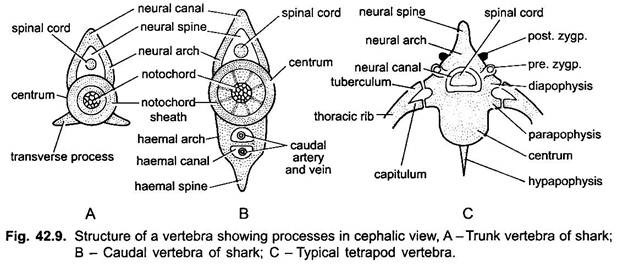
A typical vertebra has a cylindrical body or centrum which encloses or replaces embryonic notochord. Above the centrum is a neural arch enclosing a neural canal through which the nerve or spinal cord passes. The neural arch is produced dorsally into a neural spine. In the caudal region of fishes, below the centrum is a haemal arch enclosing a haemal canal through which the caudal artery and vein pass. The haemal arch is produced below into a haemal spine.
Types of Processes:
Various kinds of projections or apophyses arise from a vertebra.
They are:
(i) Zygapophyses present over anterior and posterior faces of neural arch are articulations between successive vertebrae. Prezygapophyses are anterior projections facing upwards and postzygapophyses are posterior projections facing downward,
(ii) Transverse processes- Lateral transverse processes arise from centrum,
(iii) Diapophyses arising from the neural arch or the centrum and serve for attachment of the upper head (tuberculum) of a two-headed (bicipital) rib,
(iv) Parapophyses arise laterally from the centrum for attachment of the ventral head (capitulum) of a bicipital rib,
(v) Basapophyses are ventro-lateral processes of the centrum or haemal arch, they are remnants of a haemal arch or articulate with the haemal arch when present,
(vi) Pleurapophyses are lateral projections to which short ribs are fused,
(vii) Hypapophysis is a mid-ventral projection from the centrum.
Development of Vertebrae:
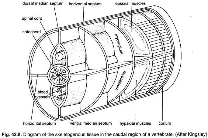
Mesenchyme cells are produced from metameric sclerotomes. A sclerotome becomes differentiated into a dense posterior or caudal half and a less dense anterior or cranial half. The caudal and cranial halves separate, then the caudal half of each sclerotome fuses with the cranial half of the following sclerotome. Thus, definite sclerotomes are formed and they come to lie inter-segmentally alternating with myotomes.
This is an adaptation by which each myotome will become connected with two successive vertebrae and their ribs, by this arrangement movement of the vertebral column occurs. The mesenchyme cells of the definite sclerotomes spread to form a skeletogenous layer around the notochord and nerve cord.
Arcualia:
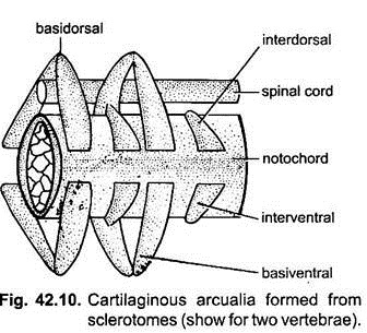
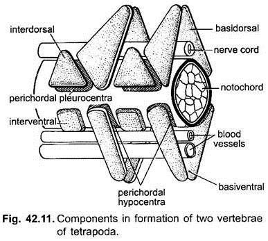
Four pairs of cartilages, called arcualia are laid down in each sclerotome lying on the two sides of the notochord. The dorsal arcualia are two interdorsals and two basidorsals, the ventral arcualia are two interventrals and two basiventrals.
The basidorsals and basiventrals are derived from the caudal half of the sclerotome and will form the anterior parts of a vertebra, while the interdorsals and interventrals arise from the cranial half of the sclerotome and will form the posterior parts of a vertebra. Generally the basidorsals and basiventrals are larger than the other arcualia.
The two basidorsals extend above the nerve cord and unite to form a neural arch.
1. Formation of Centrum:
The basiventrals, interdorsals, and interventrals form a centrum around the notochord. In the tail region the basiventrals also extend below to form a haemal arch.
2. There is another method of formation of centra, in some elasmobranchs cells from the basidorsals and basiventrals penetrate and spread into the secondary notochordal sheath and secrete cartilage to form a ring-shaped centrum around the constricted notochord, such a centrum is called a chordal centrum.
But in other elasmobranchs, bony fishes, and tetrapoda, the arcualia grow around the notochord outside the notochordal sheaths to form a centrum known as a perichordal centrum which later grows and obliterates the notochord. In both cases the centrum becomes fused to the neural and haemal arches.
3. The centrum is not always formed from arcualia only, there is another method of centrum formation. The mesenchyme lying between the definite sclerotomes and notochord, internal to the arcualia, is called the perichordal mesenchyme.
In tetrapoda the perichordal mesenchyme forms four embryonic cartilages; they are a pair of ventral hypocentra and a pair of dorsolateral pleurocentra. The hypocentrum or intercentrum incorporates the basiventrals and the pleurocentrum incorporates the interdorsals forming large pieces.
The pleurocentrum piece increases and the hypocentrum piece decreases to form the centrum of amniotes, but in amphibians the hypocentrum forms the centrum, while the pleurocentrum decreases or disappears.
Types of Centrum:
The parts forming a centrum are not always homologous and different lines of evolution are seen.
1. In crossopterygians and some fossil amphibians the two hypocentra fuses to form a large piece incomplete dorsally, but the two pleurocentra remain separate, the neural arch rests on the hypocentrum and pleurocentra, such a vertebra is rachitomous.
2. In some other extinct amphibia the hypocentrum and pleurocentrum are equal, the former is anterior and the latter posterior and each forms a disc-like centrum, but on both there is single neural arch, this is an embolomerous vertebra, it was evolved from a rachitomous vertebra.
3. On the other hand, the rachitomous vertebra gave rise to a stereospondylous vertebra of modern amphibians in which the hypocentrum enlarges to form a centrum and the pleurocentrum disappears.
4. The embolomerous vertebra gave rise to gastrocentrous vertebra of reptiles and mammals in which the pleurocentrum enlarges to form the centrum, while the hypocentrum is reduced to a small piece (reptiles) or it is changed to form an intervertebral disc (mammals).
Generally there is one vertebra per segment, this is known as monospondyly. In some cases the arcualia form two centra and only one neural arch in one segment, this is known as diplospondyly. It is found in the tail of some fishes (Amia), some extinct amphibians, and some lizards. A second type of diplospondyly is seen in the tail region of some elasmobranchs in which each segment has two complete vertebrae with two centra, two neural arches, and two haemal arches.
Types of Centra and Vertebrae:
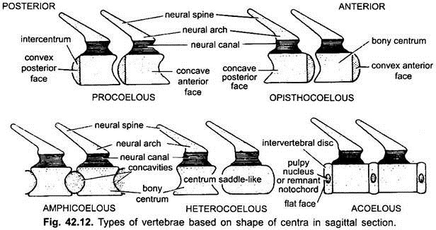
The centra of vertebra are placed end to end in a row, the shape of the ends of centra is of importance for articulation. There are six principal shapes of centra.
1. Amphicoelous:
Vertebra has its centrum concave at both ends. This is the most primitive type and is found in nearly all fishes.
2. Procoelous:
Vertebra has its centrum concave at the anterior end and convex at the posterior end. It is found in frogs and most reptiles.
3. Opisthocoelous:
Vertebra has a centrum convex at the anterior end and concave posteriorly. It is not characteristic of any group but occurs in all classes.
4. Heterocoelous:
Vertebra has the centrum saddle-shaped transversely. The anterior end is convex dorsoventrally and concave sideways, while the posterior end is opposite of the anterior end. This is the most specialised vertebra and is found in neck region of birds.
5. Amphiplatyan or Acoelous:
Vertebra has its centrum flat at both ends and is found in mammals.
6. Biconvex:
Centrum is convex at both the ends. It is found in the 9th (sacral) vertebra of frog.
Ribs:
Ribs are cartilaginous or bony structures, which are either fused to or articulated with vertebrae. Typically there is one pair of ribs to each vertebra.
In fishes there are two kinds of ribs:
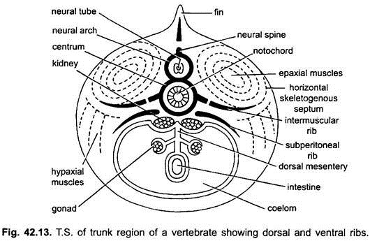
(i) Dorsal or intermuscular ribs extend from transverse processes into a skeletogenous septum between two successive myotomes.
(ii) The second kind are ventral or pleural ribs which arise from the centrum and lie between the body wall muscles and parietal peritoneum.
Most fishes have either dorsal or ventral ribs, but some have both kinds, e.g., Polypterus and many teleosts. In elasmobranchs only dorsal ribs are present, while most teleosts have only ventral ribs. In tetrapoda there is one pair of ribs to each vertebra; it is now believed that they represent the ventral ribs of fishes.
All ribs are short in tetrapoda, except those in the thorax. In amniotes the ribs of the thoracic region unite ventrally with the sternum and dorsally articulate with the vertebrae by two heads. In these two-headed or bicipital ribs the tuberculum or upper head articulates with a process of the neural arch called diapophysis, while the capitulum or lower head articulates with a process of the centrum called parapophysis (Fig. 42.9).
In the cervical region where ribs are fused with vertebrae, the space between the two heads of a rib is a vertebrarterial canal through which a nerve and blood vessels pass.
Abdominal ribs are also called gastralia or ventral ribs. There are V-shaped dermal bones in the ventral region of the abdomen extending postero-dorsally with their apices pointing in front. Gastralia were present in some fossil amphibians, in Sphenodon and crocodilians. They are dermal elements, while true ribs are endoskeleton structures.
