
In this article we will discuss about:- 1. Classification of Protozoa 2. Diagnostic Features of Protozoa 3. Scheme of Classification 4. Systematic Resume 5. Evolution.
Contents:
- Classification of Protozoa
- Diagnostic Features of Protozoa
- Scheme of Classification of Protozoa
- Systematic Resume of Protozoa
- Evolution of Protozoa
1. Classification of Protozoa:
Until the early 1970’s, all living organisms were allocated one or the other of two kingdoms — Plant or Animal. Whether an organism was uni- or multi-cellular, or whether prokaryotic or eukaryotic, were not considered relevant to this fundamental division of life. Therefore, under kingdom animal, the multicellular animals comprised the metazoa while the unicellular, the protozoa.
ADVERTISEMENTS:
Such a two-kingdom system suffers from a number of drawbacks. To name a few, it does not reflect any real phylogenetic dichotomy and the two categories are not really distinguishable. In the case of euglenoid flagellates, they have been considered under both animals and plants.
The two kingdom system is thus not a practical working scheme. R. H. Whittaker in 1969 had divided all living things into five kingdoms — Monera, Protista, Plantae, Fungi and Animalia.
While Monera is specialized at the subcellular or biochemical level, the higher organisms (Plantae, Fungi and Animalia) are specialized at the tissue level of organization. Between these two levels, the Protista is specialized at the cellular level.
Protozoa, the name coined by Goldfuss (1820), containing about 80,000 species, certainly belong to Protista that exhibits animal like mode of nutrition (Phagocytic hetero-trophy). They have been assigned the sub- kingdom category. However, according to some (Barnes et al, 1993), ‘Protozoa’ has for some time ceased to be regarded as a valid or useful grouping of organisms.
ADVERTISEMENTS:
Etymology:
Greek: protos, first; zoon, animal.
2. Diagnostic Features of Protozoa:
a. Protozoans are primitive animals with unicellular level of eukaryotic organization.
ADVERTISEMENTS:
b. Most protozoans are solitary individuals and a number of them are colonial.
c. They exploit all types of habitat and may be free-living, commensal, mutualistic and parasitic.
d. They are usually microscopic animals possessing typical membrane-bound cellular organelles.
e. They range in size from 1µm (Micro- monas) to 0.25 m (some giant, benthic marine amoeba), most being in between 5 and 250 µm in diameter.
f. Protozoan body is usually bounded only by the cell membrane. In some it may form the pellicle and in a few it is provided with simple to elaborate shells.
g. Most protozoa have a single, vesicular nucleus, while a few are multinucleate.
h. Protozoan locomotory organelles include flagella, cilia or flowing extensions of the body called pseudopodia.
i. Nutrition may be holozoic, holophytic, saprophytic or parasitic. With or without definite oral and anal apertures.
j. Both aerobic and anaerobic mode of respiration are present.
ADVERTISEMENTS:
k. Excretion either through the general body surface or through contractile vacuoles, the latter also serves for osmoregulation.
l. Reproduction commonly performed by asexual fission (both binary and multiple). In some forms sexual reproduction may occur either by conjugation or fusion of gametes.
3. Scheme of Classification of Protozoa:
Protozoans, depending upon its size, habit, habitat or depending upon its locomotory organs, had been classified differently by various authors, like Hyman (1940), Parker and Haswell (1949), Honiberg et al. (1964). The classification of Protozoa by **Levin et al. (1980) is the recent one and is widely accepted by modern taxonomists. Hence, in this text we are following the scheme of Levin et al. (1980).

4. Systematic Resume of Protozoa:
a. Phylum Sarcomastigophora:
i. Nucleus is of one type, except in heterokaryotic Foraminifera.
ii. Locomotory organs are either pseudo- podia or flagella or both.
iii. Reproduction asexually, but when sexually it is essentially by syngamy.
iv. This phylum includes three sub- phyla — Mastigophora, Opalinata and Sarcodina.
Examples:
Mastigophora — Trypanosoma, Euglena (Fig. 1.1A), Volvox, Giardia (Fig. 1.1D), Leishmania, Crypto-monas.
Opalinata — Opalina (Fig. 1.1B), Protoopalina (Fig. 1.1C).
Sarcodina — Amoeba (Fig. 1.6), Entamoeba (Fig. 1.1E), Elphidium (Fig. 1.1 F), Euglypha.

b. Phylum Labyrinthomorpha:
i. Mostly inhabitants of marine and estuarine water.
ii. Trophic stage having ectoplasmic network, with spindle shaped or spherical, non-amoeboid cells.
iii. In some genera, however, amoeboid cells move within the cytoplasmic network by gliding.
iv. Unique cell-surface organelle, associated with ectoplasmic network.
v. Saprophytic and parasitic on algae.
vi. Zoospores are produced by most species.
Examples:
Labyrinthula, Labyrinthomyxa (Fig. 1.2).

c. Phylum Apicomplexa:
i. All species are parasitic in nature.
ii. Anterior part of the body forms apical complex.
iii. This apical complex is made up of polar rings, rhoptries, micronemes, conoid and subpellicular microtubules.
iv. Microspores generally present at some stage.
v. They reproduce sexually by syngamy.
Examples:
Monocystis, Gregarina (Fig. 1.3), Plasmodium, Babesia, Perkinsus.

d. Phylum Microspora:
i. Obligatory intracellular parasites found in nearly all major animal groups.
ii. Spores unicellular, each with imperforate wall, containing one uninucleate or di-nucleate sporoplasm.
iii. Spore is with simple or complex extrusion apparatus associated with polar tube and polar cap.
iv. Mitochondria absent in spores.
v. Usually dimorphic in speculation sequence.
Examples:
Nosema (Fig. 1.4), Burkea, Hessea, Candospora.

e. Phylum Ascetospora:
i. All are parasitic.
ii. In most of the cases spores are multicellular.
iii. Spores with one or more sporoplasm.
iv. Spores without polar capsules or polar filaments.
Examples:
Haplosporidium, Coelosporidium, Paramyxa, Marteilia.
f. Phylum Myxozoa:
i. All species are parasitic.
ii. Spores are of multicellular origin, with one or more polar capsules and sporoplasms.
iii. Each spore with 1, 2 or 3 (rarely more) valves.
Examples:
Myxidium, Myxobolus, Trilospora, Triactinomyxon.
g. Phylum Ciliophora:
i. Most of the species are free living, quite a number are commensal, some truly parasitic and a large number are found as symphorionts on variety of hosts.
ii. Simple cilia or compound ciliary organelles are present in at least one stage of life cycle.
iii. Subpellicular cilia is present even when surface cilia is absent.
iv. Nuclei are of two types.
v. Presence of typical contractile vacuole.
vi. Nutrition heterotrophic.
vii. Asexual reproduction by transverse binary fission, basically homothetogenic and generally parakinetal, but budding and multiple fission also occur.
viii. Sexual reproduction involves conjugation, autogamy and cytogamy.
Examples:
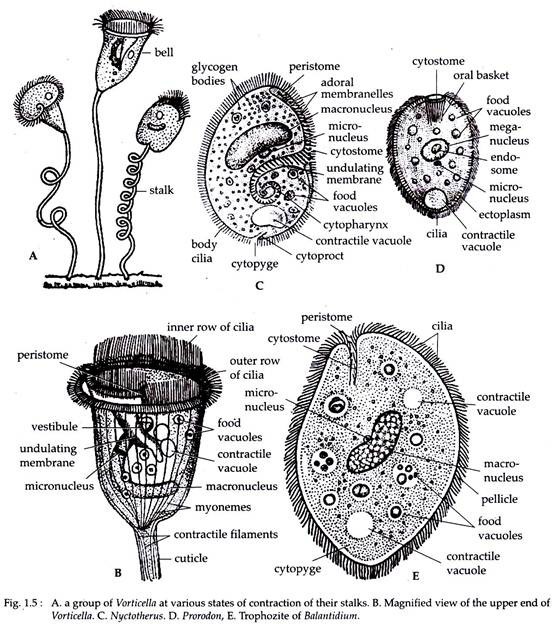
Paramoecium (Fig. 1.15), Balantidiuma (Fig. 1.5E), Vorticella (Fig. 1.5A and B), Nyctotherus (Fig. 1.5C), Tetrahymena, Nassula, Podophrya, Prorodon (Fig. 1.5D).
5. Evolution of Protozoa:
The evolution of protozoa is the history of eukaryote cell and the different stages in that history has been well reflected by the living ones present today. The earliest protists were probably amoeboid forms capturing food particles by phagocytosis. In one such engulfment of a symbiotic prokaryotic organism, some amoeboid probably had acquired mitochondria.
Flagella may also have been acquired early, both prior or later to the acquirement of mitochondria. This has been proved as some living flagellates lack mitochondria (Giardia). Many living flagellates possess chloroplasts which also may have been obtained by the process of phagocytosis of prokaryotic symbionts by some early flagellated eukaryotes.
This can be seen as some living flagellates have retained their ancestral phagocytosis nature along with photosynthetic capabilities (some dinoflagellates), while others are solely photoautotrophs (Euglena, Volvox etc.). Contributing to this confusion of protozoan evolution are some species that have lost their chloroplasts and are secondarily heterotrophic (Peranema, Leishmania, Trypanosoma etc.).
Some even have lost their flagella and are secondarily amoeboid. Therefore, some amoebas are probably more closely related to some flagellates than they are to other amoeboid groups as they possess flagellated gametes in their life cycle (Discorbis). However, only the ciliates are likely to be a natural group.