In this article we will discuss about the structure and life history of trypanosoma, explained with help of suitable diagrams.
Structure of Trypanosoma:
1. Trypanosoma is a polymorphic animal having a flattened, elongate-fusiform body.
2. The anterior end is pointed and the posterior end blunt.
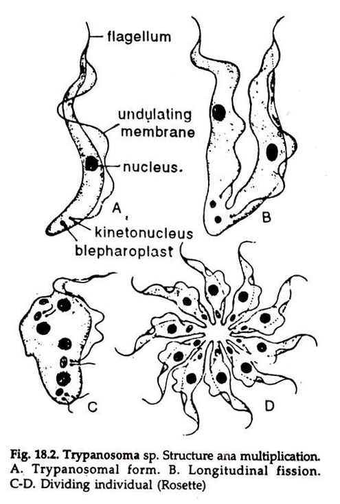
3. It measures 15 to 60 µm in length and 1.5 to 3.5 µm in width (Fig. 18.2A).
4. A strong, tough, elastic pellicle encloses the body. The pellicle is lined by a layer of ectoplasm and the central mass is endoplasm.
5. A large vesicular nucleus is present in the centre of the body.
6. Behind the nucleus and near the posterior end of the body is a spherical or rod- shaped or disc-shaped parabasal body or kinetoplast. A small blepharoplast is present in front of it. The two are connected by a delicate rhizoplast.
7. A flagellum arises from the posterior end of the blepharoplast and extends forward, towards the anterior end along the outer margin of the body, being connected to it by an undulating membrane.
ADVERTISEMENTS:
8. A small portion of the flagellum extends freely beyond the anterior end.
9. The contractile vacuoles are absent.
10. Volentine granules in the form of refractile bodies are scattered in the endoplasm.
Trypanosoma are polymorphic and occur in four morphologically different forms:
ADVERTISEMENTS:
a. Leishmanial stage:
Without flagellum and with anteriorly placed kinetoplast.
b. Leptomonad stage:
Elongated body with a broad anterior end, a free flagellum and an anteriorly placed kinetoplast.
c. Crithidial stage:
Kinetoplast and flagellum situated in front, anterior to the nucleus.
d. Trypanosome stage:
Kinetoplast situated far behind the nucleus near the posterior end of the body.
Life History of Trypanosoma:
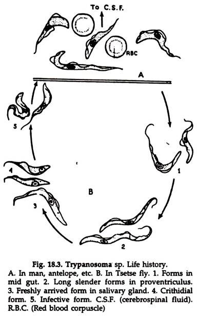
1. Trypanosoma completes its life cycle in two hosts (Fig. 18.3). The definitive host is a vertebrate and the intermediate or secondary host is an invertebrate, usually a Tsetse fly, Glossina sp.
2. In the vertebrate host, trypanosomes usually live in the blood or cerebrospinal fluid.
Inoculation:
3. With the bite of Tsetse fly, the parasites enter the blood stream of man in the metacyclic form. In some cases the parasites may reach cerebrospinal fluid from the blood.
Vertebrate Host:
4. In the blood stream the parasites multiply by binary fission (Fig. 18.2B).
And is represented in three morphological forms:
i. Slender form:
With a large flagellum.
ii. Intermediate form:
With a short flagellum.
iii. Stumpy form:
With a broad body devoid of a flagellum.
Secondary Host:
5. The Tsetse fly (Glossina palpalis) sucks the trypanosomes along with host’s blood. The parasites remain unchanged in the midgut for a few hours.
6. In favourable conditions the parasites multiply by repeated binary fission (Fig. 18.2C, D) and after about two weeks numerous slender forms appear. These gradually ascend the foregut.
7. By the end of the third week some of these slender forms reach the salivary gland through the labial cavity and hypo pharynx.
8. In the salivary glands the parasites change into crithidal stage.
9. They multiply rapidly for about 2-3 days and finally metacyclic trypanosomes are formed.
10. These are the infective forms and are to be transferred to the primary host.

