In this article we will discuss about the life history of entamoeba histolytica, explained with the help of suitable diagrams.
Structure of Entamoeba Histolytica:
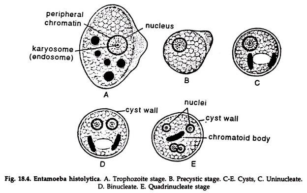
The amoeba has three stages in its life cycle, viz. the trophozoite stage, the precystic stage and the cystic stage.
A. The trophozoite amoeba:
ADVERTISEMENTS:
This is the growing or feeding stage of the parasite having the following features:
1. The trophozoite is somewhat elongated (Fig. 18.4A); measuring 18 to 40 µm, but the shape is not fixed as the animal constantly changes its contour.
2. The cytoplasm is divisible into a clear translucent ectoplasm and a granular endoplasm.
3. The nucleus is spherical and varies from 4 to 6 µm in size.
ADVERTISEMENTS:
a. The nucleus is -surrounded by a delicate nuclear membrane lined with a single layer of chromatin granules.
b. The centre of the nucleus is occupied by a karyosome surrounded by a clear halo.
c. The space between the membrane and the karyosome is traversed by fine threads arranged radially in the form of spokes of a wheel.
4. Ingested blood cells are found in the cytoplasm.
ADVERTISEMENTS:
Encystation:
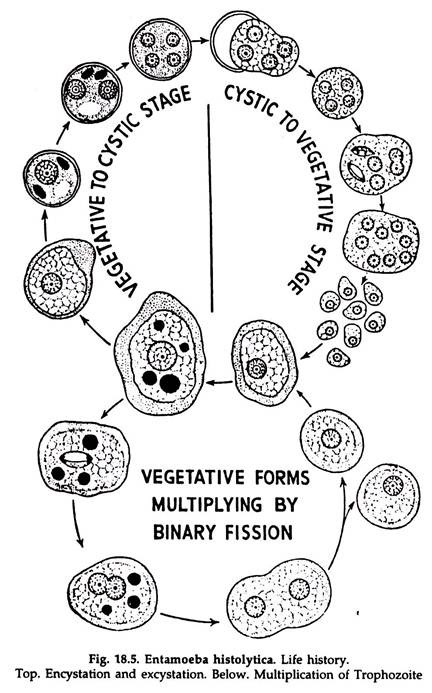
In some cases, the trophozoites are discharged into the lumen from the intestinal wall and are transformed into precystic forms, which produce cystic forms. This phenomenon is known as encystation and leads to the production of a different strain.
B. Precystic amoeba:
1. This is slightly ovoid with a short and blunt pseudopodium projecting from one side (Fig. 18.4B).
2. Size varies from 10 to 20 µm.
3. The motility ceases and ingested particles are extruded.
C. Cystic amoeba:
1. The parasite becomes rounded and surrounds itself by a highly refractile cyst wall (Fig. 18.4C-E).
2. Size varies from 7 to 20 µm.
ADVERTISEMENTS:
3. Contains one or more chromatid bodies with round ends.
4. The cyst is uninucleate at the beginning, but undergoes double fission so that four nuclei are produced. The quadrinucleate stage is considered as the mature cyst.
Excystation:
The liberation of the parasite from the cyst is known as excystation. The mature cysts are infective forms. They reach the alimentary canal with contaminated food and drink.
The cyst wall is digested by the action of trypsin in the intestine. When the cyst reaches the iliocaecal junction, particularly the caecum, vigorous amoeboid movement starts in the cytoplasm and a rent appears in the cyst wall. The parasite comes out through the rent.
The four nuclei now divide into eight, the cytoplasm also undergoes division and eight minute amoebulae, also called. metacystic amoebae, are formed. The young amoebae are active and invade the intestinal tissue to reach the mucous and sub mucous layers, their normal habitat.
Reproduction in Entamoeba Histolytica:
Entamoeba reproduces (Fig. 18.5) by binary fission, the rate of multiplication being very high.
1. The nucleus slightly elongate and assumes an ovoid shape.
2. A constriction appears in the middle which grows deeper and the nucleus is divided into two by a modified type of mitosis.
3. The cytoplasmic mass elongates slightly, a constriction appears at the middle which grows deeper and the parasite is divided into two equal halves.
4. Each half receives one daughter nucleus and two individuals are thus produced.
Modes of infection in Entamoeba Histolytica:
The life cycle of Entamoeba histolytica passes only in one host, the man. Transmission from man to man is effected through faecal contamination of drinking water and vegetables directly or through the agency of flies or cockroaches.
The mature quadrinucleate cysts are infective forms. Eating of uncooked vegetables and fruits and drinking of water contaminated with infective forms of parasites lead to the infection of a new host.
Cysts can pass through the intestine of the fly or cockroach in a viable condition, and infest new hosts through contaminated food. Cooks, mess boys and food handlers are important transmitters of infection in tropical countries.

